Chen Li, Song Jiarui, Cheng Runtian, Wang Kangcheng, Liu Xiaoshuang, He Miao, Luo Tianyou
Department of Radiology, Affiliated Hospital of North Sichuan Medical College, Nanchong, China.
Department of Radiology, The First Affiliated Hospital of Chongqing Medical University, Chongqing, China.
Front Aging Neurosci. 2021 Feb 17;12:614833. doi: 10.3389/fnagi.2020.614833. eCollection 2020.
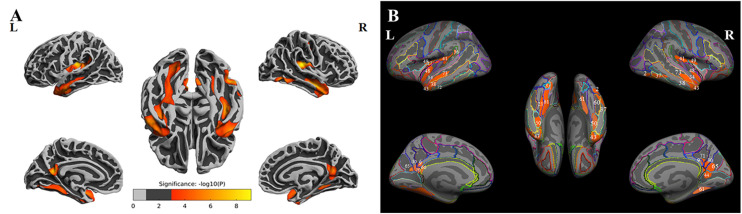
Subcortical ischemic vascular disease (SIVD) is a major cause of vascular cognitive impairment (CI) and features extensive atrophy in the cerebral cortex. We aimed to test the hypothesis that cognitive deficits in SIVD are linked to decreased cortical thickness in specific brain regions, which may constitute neuroimaging biomarkers of CI. Sixty-seven SIVD patients without (SIVD-NC, = 35) and with (SIVD-CI, = 32) CI and a group of healthy controls (HCs, = 36) underwent structural magnetic resonance imaging (MRI) and cognitive functional assessments. FreeSurfer was used to preprocess structural MRI data and to calculate and compare cortical thickness. The correlation between cortical thickness and cognitive scores was examined in SIVD patients. Significantly altered cortical thickness in the bilateral insula, middle and inferior temporal lobes, precuneus, and medial temporal lobe (MTL) was identified among the three groups ( < 0.05, Monte Carlo simulation corrected). results showed significantly decreased thickness in the bilateral insula and temporal lobe in SIVD-NC and SIVD-CI patients compared with HCs. However, the areas with reduced cortical thickness were larger in SIVD-CI than SIVD-NC patients. SIVD-CI patients had significantly reduced thickness in the bilateral precuneus and left MTL (Bonferroni corrected) compared with SIVD-NC patients when we extracted the mean thickness for each region of interest. In SIVD patients, the thicknesses of the left MTL and bilateral precuneus were positively correlated with immediate recall in the memory test. SIVD might lead to extensive cerebral cortical atrophy, while atrophy in the MTL and precuneus might be associated with memory deficits.
皮质下缺血性血管疾病(SIVD)是血管性认知障碍(CI)的主要原因,其特征是大脑皮质广泛萎缩。我们旨在验证以下假设:SIVD中的认知缺陷与特定脑区皮质厚度降低有关,这些脑区可能构成CI的神经影像学生物标志物。67例无CI(SIVD-NC,n = 35)和有CI(SIVD-CI,n = 32)的SIVD患者以及一组健康对照(HCs,n = 36)接受了结构磁共振成像(MRI)和认知功能评估。使用FreeSurfer对结构MRI数据进行预处理,并计算和比较皮质厚度。在SIVD患者中检查皮质厚度与认知评分之间的相关性。三组之间双侧岛叶、颞叶中下部、楔前叶和内侧颞叶(MTL)的皮质厚度有显著改变(P < 0.05,经蒙特卡罗模拟校正)。结果显示,与HCs相比,SIVD-NC和SIVD-CI患者的双侧岛叶和颞叶厚度显著降低。然而,SIVD-CI患者皮质厚度降低的区域比SIVD-NC患者更大。当我们提取每个感兴趣区域的平均厚度时,与SIVD-NC患者相比,SIVD-CI患者的双侧楔前叶和左侧MTL厚度显著降低(经Bonferroni校正)。在SIVD患者中,左侧MTL和双侧楔前叶的厚度与记忆测试中的即时回忆呈正相关。SIVD可能导致广泛的大脑皮质萎缩,而MTL和楔前叶的萎缩可能与记忆缺陷有关。