González-Molina Luis Alfonso, Villar-Vesga Juan, Henao-Restrepo Julián, Villegas Andrés, Lopera Francisco, Cardona-Gómez Gloria Patricia, Posada-Duque Rafael
Área de Neurobiología Celular y Molecular, Grupo de Neurociencias de Antioquia, Universidad de Antioquia, Medellin, Colombia.
Facultad de Ciencias Exactas y Naturales, Instituto de Biología, Universidad de Antioquia, Medellin, Colombia.
Front Aging Neurosci. 2021 Feb 19;13:593927. doi: 10.3389/fnagi.2021.593927. eCollection 2021.
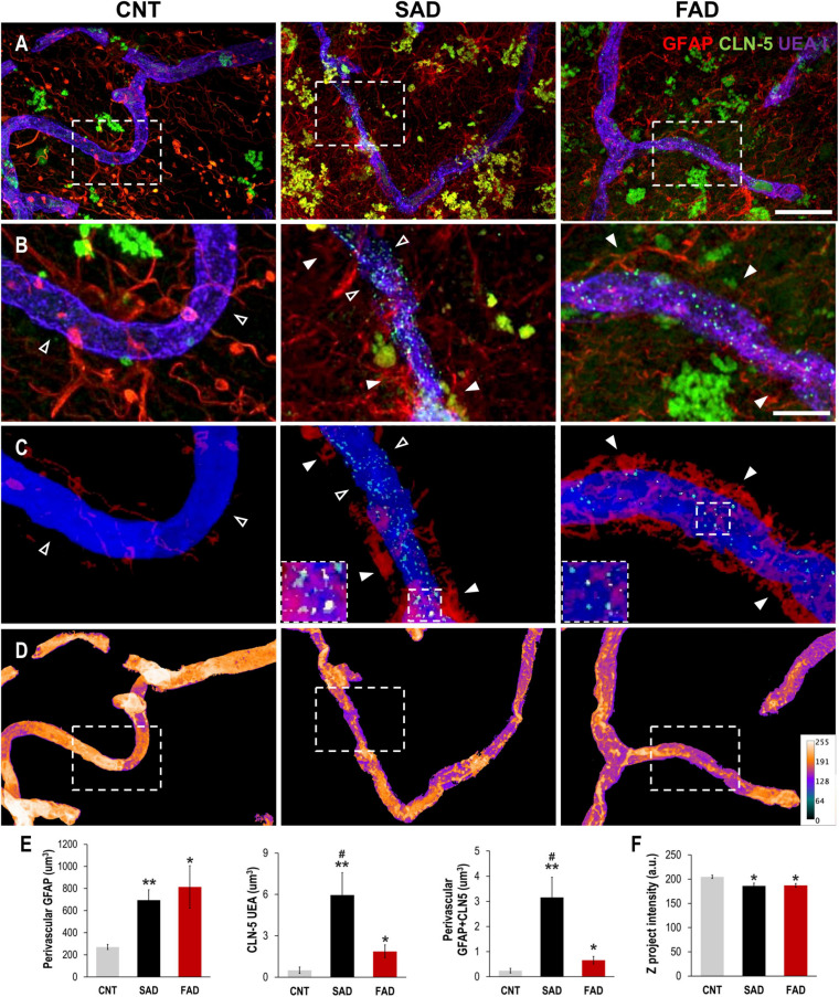
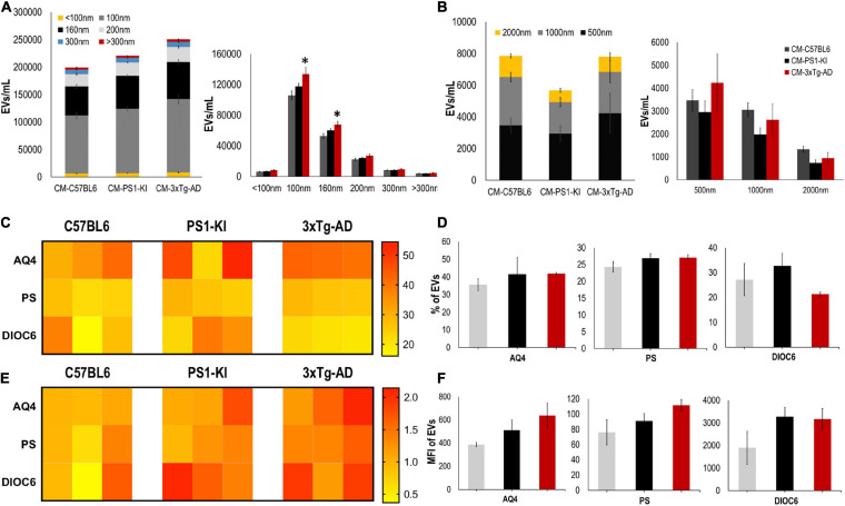
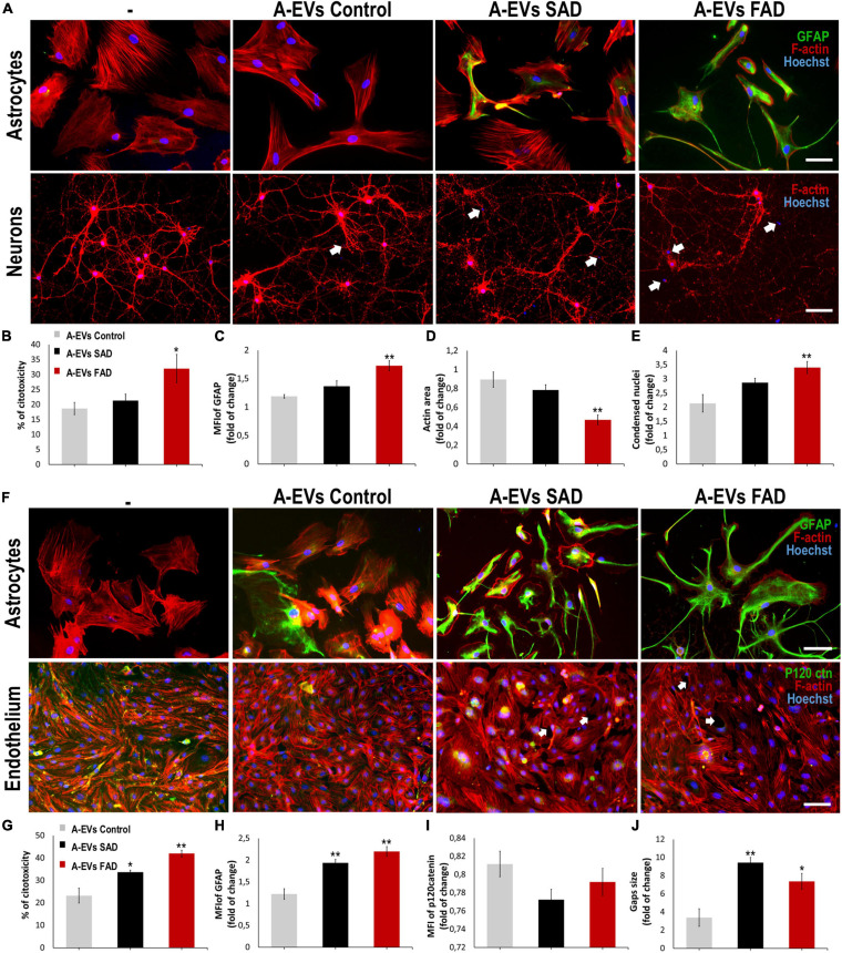
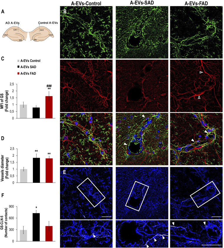
Astrocytes are specialized glial cells that are essential components of the neurovascular unit (NVU) and are involved in neurodevelopment, brain maintenance and repair, and neurodegeneration. Astrocytes mediate these processes by releasing cellular mediators such as extracellular vesicles (EVs). EVs are vehicles of cell-cell communication and have been proposed as mediators of damage in AD. However, the transcellular mechanism by which Alzheimer disease (AD) astrocytes impair the function of NVU components is poorly understood. Therefore, we evaluated the effects of adult PS1-KI and 3xTg-AD astrocyte conditioned media (CM) and EVs on NVU components (neuroglia and endothelium) . Additionally, SAD and FAD astrocyte-derived EVs (A-EVs) were characterized, and we evaluated their effects on NVU in cocultured cells and on intrahippocampal CA1 cells . Surprisingly, cultured 3xTg-AD astrocytes showed increased glial fibrillary acidic protein (GFAP) reactivity compared to PS1-KI astrocytes, which denotes astrocytic hyperreactivity. CM from adult mice 3xTg-AD astrocytes increased cell-cell gaps between endothelial cells, filopodia-like dendritic protrusions in neurons and neuronal and endothelial cell death. 3xTg-AD A-EVs induced neurotoxicity and increased astrocyte GFAP reactivity. Cultured human astrocytes from AD patients also increased GFAP reactivity and EVs release. No differences in the size or number of A-EVs were detected between AD and control samples; however, both SAD and FAD A-EVs showed increased expression of the surface marker aquaporin 4. A-EVs induced cytotoxicity and astrocyte hyperactivation: specifically, FAD A-EVs induced neuroglial cytotoxicity and increased gaps between the endothelium, while SAD A-EVs mainly altered the endothelium. Similarly, both AD A-EVs increased astrocyte GS reactivity and vascular deterioration . We associated this finding with perivascular reactive astrocytes and vascular deterioration in the human AD brain. In summary, these results suggest that AD A-EVs impair neuroglial and vascular components.
星形胶质细胞是一种特殊的神经胶质细胞,是神经血管单元(NVU)的重要组成部分,参与神经发育、大脑维持与修复以及神经退行性变。星形胶质细胞通过释放细胞介质如细胞外囊泡(EVs)来介导这些过程。EVs是细胞间通讯的载体,被认为是阿尔茨海默病(AD)中损伤的介质。然而,AD星形胶质细胞损害NVU组分功能的跨细胞机制尚不清楚。因此,我们评估了成年PS1-KI和3xTg-AD星形胶质细胞条件培养基(CM)及EVs对NVU组分(神经胶质和内皮)的影响。此外,对散发性AD(SAD)和家族性AD(FAD)星形胶质细胞衍生的EVs(A-EVs)进行了表征,并评估了它们对共培养细胞中NVU以及海马CA1区内细胞的影响。令人惊讶的是,与PS1-KI星形胶质细胞相比,培养的3xTg-AD星形胶质细胞显示出更高的胶质纤维酸性蛋白(GFAP)反应性,这表明星形胶质细胞反应过度。成年小鼠3xTg-AD星形胶质细胞的CM增加了内皮细胞之间的细胞间隙、神经元中丝状伪足样树突状突起以及神经元和内皮细胞死亡。3xTg-AD A-EVs诱导神经毒性并增加星形胶质细胞GFAP反应性。来自AD患者的培养人星形胶质细胞也增加了GFAP反应性和EVs释放。在AD和对照样本之间未检测到A-EVs大小或数量的差异;然而,SAD和FAD的A-EVs均显示水通道蛋白4表面标志物表达增加。A-EVs诱导细胞毒性和星形胶质细胞过度激活:具体而言,FAD A-EVs诱导神经胶质细胞毒性并增加内皮之间的间隙,而SAD A-EVs主要改变内皮。同样,两种AD A-EVs均增加星形胶质细胞谷氨酰胺合成酶(GS)反应性和血管退化。我们将这一发现与人类AD大脑中血管周围反应性星形胶质细胞和血管退化联系起来。总之,这些结果表明AD A-EVs损害神经胶质和血管组分。