• 文献检索
  • 文档翻译
  • 深度研究
  • 学术资讯
  • Suppr Zotero 插件Zotero 插件
  • 邀请有礼
  • 套餐&价格
  • 历史记录
应用&插件
Suppr Zotero 插件Zotero 插件浏览器插件Mac 客户端Windows 客户端微信小程序
定价
高级版会员购买积分包购买API积分包
服务
文献检索文档翻译深度研究API 文档MCP 服务
关于我们
关于 Suppr公司介绍联系我们用户协议隐私条款
关注我们

Suppr 超能文献

核心技术专利:CN118964589B侵权必究
粤ICP备2023148730 号-1Suppr @ 2026

文献检索

告别复杂PubMed语法,用中文像聊天一样搜索,搜遍4000万医学文献。AI智能推荐,让科研检索更轻松。

立即免费搜索

文件翻译

保留排版,准确专业,支持PDF/Word/PPT等文件格式,支持 12+语言互译。

免费翻译文档

深度研究

AI帮你快速写综述,25分钟生成高质量综述,智能提取关键信息,辅助科研写作。

立即免费体验

基于卷积神经网络的肾肿瘤超声造影图像自动分割及影像组学特征可靠性研究

CNN-based automatic segmentations and radiomics feature reliability on contrast-enhanced ultrasound images for renal tumors.

作者信息

Yang Yin, Chen Fei, Liang Hongmei, Bai Yun, Wang Zhen, Zhao Lei, Ma Sai, Niu Qinghua, Li Fan, Xie Tianwu, Cai Yingyu

机构信息

Department of Ultrasound, Shanghai General Hospital, Shanghai Jiao Tong University School of Medicine, Shanghai, China.

Department of Pediatrics, Jiahui International Hospital, Shanghai, China.

出版信息

Front Oncol. 2023 Jun 2;13:1166988. doi: 10.3389/fonc.2023.1166988. eCollection 2023.

DOI:10.3389/fonc.2023.1166988
PMID:37333811
原文链接:https://pmc.ncbi.nlm.nih.gov/articles/PMC10272725/
Abstract

OBJECTIVE

To investigate the feasibility and efficiency of automatic segmentation of contrast-enhanced ultrasound (CEUS) images in renal tumors by convolutional neural network (CNN) based models and their further application in radiomic analysis.

MATERIALS AND METHODS

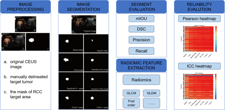
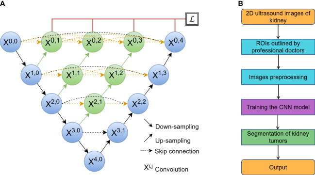
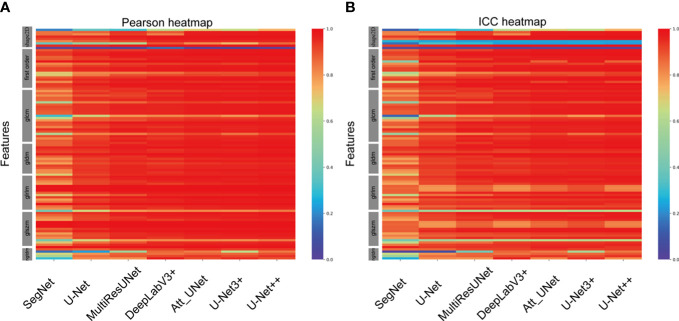
From 94 pathologically confirmed renal tumor cases, 3355 CEUS images were extracted and randomly divided into training set (3020 images) and test set (335 images). According to the histological subtypes of renal cell carcinoma, the test set was further split into clear cell renal cell carcinoma (ccRCC) set (225 images), renal angiomyolipoma (AML) set (77 images) and set of other subtypes (33 images). Manual segmentation was the gold standard and serves as ground truth. Seven CNN-based models including DeepLabV3+, UNet, UNet++, UNet3+, SegNet, MultilResUNet and Attention UNet were used for automatic segmentation. Python 3.7.0 and Pyradiomics package 3.0.1 were used for radiomic feature extraction. Performance of all approaches was evaluated by the metrics of mean intersection over union (mIOU), dice similarity coefficient (DSC), precision, and recall. Reliability and reproducibility of radiomics features were evaluated by the Pearson coefficient and the intraclass correlation coefficient (ICC).

RESULTS

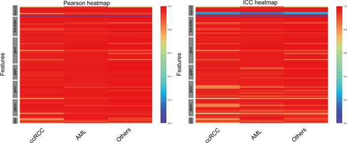
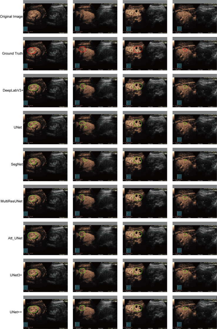
All seven CNN-based models achieved good performance with the mIOU, DSC, precision and recall ranging between 81.97%-93.04%, 78.67%-92.70%, 93.92%-97.56%, and 85.29%-95.17%, respectively. The average Pearson coefficients ranged from 0.81 to 0.95, and the average ICCs ranged from 0.77 to 0.92. The UNet++ model showed the best performance with the mIOU, DSC, precision and recall of 93.04%, 92.70%, 97.43% and 95.17%, respectively. For ccRCC, AML and other subtypes, the reliability and reproducibility of radiomic analysis derived from automatically segmented CEUS images were excellent, with the average Pearson coefficients of 0.95, 0.96 and 0.96, and the average ICCs for different subtypes were 0.91, 0.93 and 0.94, respectively.

CONCLUSION

This retrospective single-center study showed that the CNN-based models had good performance on automatic segmentation of CEUS images for renal tumors, especially the UNet++ model. The radiomics features extracted from automatically segmented CEUS images were feasible and reliable, and further validation by multi-center research is necessary.

摘要

目的

探讨基于卷积神经网络(CNN)的模型对肾肿瘤超声造影(CEUS)图像进行自动分割的可行性和效率,及其在放射组学分析中的进一步应用。

材料与方法

从94例经病理证实的肾肿瘤病例中提取3355幅CEUS图像,并随机分为训练集(3020幅图像)和测试集(335幅图像)。根据肾细胞癌的组织学亚型,测试集进一步分为透明细胞肾细胞癌(ccRCC)组(225幅图像)、肾血管平滑肌脂肪瘤(AML)组(77幅图像)和其他亚型组(33幅图像)。手动分割为金标准并作为真实对照。使用包括DeepLabV3 +、UNet、UNet ++、UNet3 +、SegNet、MultilResUNet和Attention UNet在内的7种基于CNN的模型进行自动分割。使用Python 3.7.0和Pyradiomics软件包3.0.1进行放射组学特征提取。所有方法的性能通过平均交并比(mIOU)、骰子相似系数(DSC)、精度和召回率指标进行评估。通过Pearson系数和组内相关系数(ICC)评估放射组学特征的可靠性和可重复性。

结果

所有7种基于CNN的模型均取得了良好的性能,mIOU、DSC、精度和召回率分别在81.97% - 93.04%、78.67% - 92.70%、93.92% - 97.56%和85.29% - 95.17%之间。平均Pearson系数在0.81至0.95之间,平均ICC在0.77至0.92之间。UNet ++模型表现最佳,mIOU、DSC、精度和召回率分别为93.04%、92.70%、97.43%和95.17%。对于ccRCC、AML和其他亚型,从自动分割的CEUS图像中得出的放射组学分析的可靠性和可重复性极佳,平均Pearson系数分别为0.95、0.96和0.96,不同亚型的平均ICC分别为0.91、0.93和0.94。

结论

这项回顾性单中心研究表明,基于CNN的模型在肾肿瘤CEUS图像的自动分割方面表现良好,尤其是UNet ++模型。从自动分割的CEUS图像中提取的放射组学特征是可行且可靠的,需要通过多中心研究进行进一步验证。

https://cdn.ncbi.nlm.nih.gov/pmc/blobs/8111/10272725/daa7155fdd44/fonc-13-1166988-g005.jpg
https://cdn.ncbi.nlm.nih.gov/pmc/blobs/8111/10272725/b5c88cb8edf7/fonc-13-1166988-g001.jpg
https://cdn.ncbi.nlm.nih.gov/pmc/blobs/8111/10272725/48db14e2af79/fonc-13-1166988-g002.jpg
https://cdn.ncbi.nlm.nih.gov/pmc/blobs/8111/10272725/25389f01d099/fonc-13-1166988-g003.jpg
https://cdn.ncbi.nlm.nih.gov/pmc/blobs/8111/10272725/bb598cd908df/fonc-13-1166988-g004.jpg
https://cdn.ncbi.nlm.nih.gov/pmc/blobs/8111/10272725/daa7155fdd44/fonc-13-1166988-g005.jpg

相似文献

1
CNN-based automatic segmentations and radiomics feature reliability on contrast-enhanced ultrasound images for renal tumors.基于卷积神经网络的肾肿瘤超声造影图像自动分割及影像组学特征可靠性研究
Front Oncol. 2023 Jun 2;13:1166988. doi: 10.3389/fonc.2023.1166988. eCollection 2023.
2
Multiple U-Net-Based Automatic Segmentations and Radiomics Feature Stability on Ultrasound Images for Patients With Ovarian Cancer.基于多U-Net的卵巢癌患者超声图像自动分割及影像组学特征稳定性研究
Front Oncol. 2021 Feb 18;10:614201. doi: 10.3389/fonc.2020.614201. eCollection 2020.
3
The Accuracy and Radiomics Feature Effects of Multiple U-net-Based Automatic Segmentation Models for Transvaginal Ultrasound Images of Cervical Cancer.基于多 U-net 的自动分割模型对宫颈癌经阴道超声图像的准确性和放射组学特征效果。
J Digit Imaging. 2022 Aug;35(4):983-992. doi: 10.1007/s10278-022-00620-z. Epub 2022 Mar 30.
4
A deep-learning approach for segmentation of liver tumors in magnetic resonance imaging using UNet+.基于 UNet+的深度学习方法在磁共振成像中对肝脏肿瘤进行分割。
BMC Cancer. 2023 Nov 3;23(1):1060. doi: 10.1186/s12885-023-11432-x.
5
Deep learning-based automatic segmentation of meningioma from T1-weighted contrast-enhanced MRI for preoperative meningioma differentiation using radiomic features.基于深度学习的 T1 加权对比增强 MRI 脑膜瘤自动分割用于术前脑膜瘤分化的放射组学特征。
BMC Med Imaging. 2024 Mar 5;24(1):56. doi: 10.1186/s12880-024-01218-3.
6
Deep learning-based recognition and segmentation of intracranial aneurysms under small sample size.小样本量下基于深度学习的颅内动脉瘤识别与分割
Front Physiol. 2022 Dec 19;13:1084202. doi: 10.3389/fphys.2022.1084202. eCollection 2022.
7
Deep learning enables automatic quantitative assessment of puborectalis muscle and urogenital hiatus in plane of minimal hiatal dimensions.深度学习可实现肛提肌和尿生殖膈在最小裂孔平面的自动定量评估。
Ultrasound Obstet Gynecol. 2019 Aug;54(2):270-275. doi: 10.1002/uog.20181. Epub 2019 Jun 26.
8
Radiomics as a measure superior to common similarity metrics for tumor segmentation performance evaluation.放射组学作为一种优于常用相似性度量的肿瘤分割性能评估方法。
J Appl Clin Med Phys. 2024 Aug;25(8):e14442. doi: 10.1002/acm2.14442. Epub 2024 Jun 23.
9
Deep Learning-Based Segmentation and Risk Stratification for Gastrointestinal Stromal Tumors in Transabdominal Ultrasound Imaging.基于深度学习的经腹超声成像中胃肠道间质瘤的分割与风险分层
J Ultrasound Med. 2024 Sep;43(9):1661-1672. doi: 10.1002/jum.16489. Epub 2024 May 31.
10
The Effects of Automatic Segmentations on Preoperative Lymph Node Status Prediction Models With Ultrasound Radiomics for Patients With Early Stage Cervical Cancer.自动分割对基于超声放射组学的早期宫颈癌患者术前淋巴结状态预测模型的影响。
Technol Cancer Res Treat. 2022 Jan-Dec;21:15330338221099396. doi: 10.1177/15330338221099396.

引用本文的文献

1
Artificial Intelligence-Augmented Advancements in the Diagnostic Challenges Within Renal Cell Carcinoma.人工智能助力肾细胞癌诊断挑战的进展
J Clin Med. 2025 Mar 26;14(7):2272. doi: 10.3390/jcm14072272.
2
Ultrasound-based radiogenomics: status, applications, and future direction.基于超声的放射基因组学:现状、应用及未来方向。
Ultrasonography. 2025 Mar;44(2):95-111. doi: 10.14366/usg.24152. Epub 2024 Dec 12.
3
The impact of BTK knockdown on lung adenocarcinoma growth and immune response.布鲁顿酪氨酸激酶(BTK)敲低对肺腺癌生长及免疫反应的影响。

本文引用的文献

1
Ultrasound Image-Based Deep Features and Radiomics for the Discrimination of Small Fat-Poor Angiomyolipoma and Small Renal Cell Carcinoma.基于超声图像的深度学习特征和放射组学在小的乏脂性血管平滑肌脂肪瘤和小肾癌鉴别诊断中的应用。
Ultrasound Med Biol. 2023 Feb;49(2):560-568. doi: 10.1016/j.ultrasmedbio.2022.10.009. Epub 2022 Nov 12.
2
Prediction of early recurrence of HCC after hepatectomy by contrast-enhanced ultrasound-based deep learning radiomics.基于超声造影的深度学习影像组学预测肝癌肝切除术后早期复发
Front Oncol. 2022 Sep 28;12:930458. doi: 10.3389/fonc.2022.930458. eCollection 2022.
3
Attention UW-Net: A fully connected model for automatic segmentation and annotation of chest X-ray.
Cancer Sci. 2025 Jun;116(6):1550-1564. doi: 10.1111/cas.16394. Epub 2024 Dec 4.
4
Segmentation of glioblastomas via 3D FusionNet.通过3D融合网络对胶质母细胞瘤进行分割
Front Oncol. 2024 Nov 15;14:1488616. doi: 10.3389/fonc.2024.1488616. eCollection 2024.
5
Automation of Wilms' tumor segmentation by artificial intelligence.人工智能在威尔姆斯瘤分割中的自动化应用。
Cancer Imaging. 2024 Jul 2;24(1):83. doi: 10.1186/s40644-024-00729-0.
注意 UW-Net:一种用于自动分割和注释胸部 X 光的全连接模型。
Comput Biol Med. 2022 Nov;150:106083. doi: 10.1016/j.compbiomed.2022.106083. Epub 2022 Sep 10.
4
Contrast-enhanced ultrasound (CEUS) of benign and malignant renal tumors: Distinguishing CEUS features differ with tumor size.增强超声(CEUS)在良恶性肾肿瘤中的应用:不同大小肿瘤的 CEUS 特征具有鉴别意义。
Cancer Med. 2023 Feb;12(3):2551-2559. doi: 10.1002/cam4.5101. Epub 2022 Sep 4.
5
A deep learning-based precision volume calculation approach for kidney and tumor segmentation on computed tomography images.基于深度学习的 CT 图像肾脏和肿瘤分割的精准体积计算方法。
Comput Methods Programs Biomed. 2022 Jun;221:106861. doi: 10.1016/j.cmpb.2022.106861. Epub 2022 May 10.
6
The Accuracy and Radiomics Feature Effects of Multiple U-net-Based Automatic Segmentation Models for Transvaginal Ultrasound Images of Cervical Cancer.基于多 U-net 的自动分割模型对宫颈癌经阴道超声图像的准确性和放射组学特征效果。
J Digit Imaging. 2022 Aug;35(4):983-992. doi: 10.1007/s10278-022-00620-z. Epub 2022 Mar 30.
7
A novel convolutional neural network for kidney ultrasound images segmentation.一种用于肾脏超声图像分割的新型卷积神经网络。
Comput Methods Programs Biomed. 2022 May;218:106712. doi: 10.1016/j.cmpb.2022.106712. Epub 2022 Feb 26.
8
Hyperechoic Renal Masses: Differentiation of Angiomyolipomas from Renal Cell Carcinomas using Tumor Size and Ultrasound Radiomics.高回声性肾肿物:应用肿瘤大小和超声放射组学对血管平滑肌脂肪瘤与肾细胞癌进行鉴别诊断。
Ultrasound Med Biol. 2022 May;48(5):887-894. doi: 10.1016/j.ultrasmedbio.2022.01.011. Epub 2022 Feb 23.
9
Exploring the Value of Radiomics Features Based on B-Mode and Contrast-Enhanced Ultrasound in Discriminating the Nature of Thyroid Nodules.基于B超和超声造影的影像组学特征在鉴别甲状腺结节性质中的价值探讨
Front Oncol. 2021 Oct 14;11:738909. doi: 10.3389/fonc.2021.738909. eCollection 2021.
10
Multiple U-Net-Based Automatic Segmentations and Radiomics Feature Stability on Ultrasound Images for Patients With Ovarian Cancer.基于多U-Net的卵巢癌患者超声图像自动分割及影像组学特征稳定性研究
Front Oncol. 2021 Feb 18;10:614201. doi: 10.3389/fonc.2020.614201. eCollection 2020.