• 文献检索
  • 文档翻译
  • 深度研究
  • 学术资讯
  • Suppr Zotero 插件Zotero 插件
  • 邀请有礼
  • 套餐&价格
  • 历史记录
应用&插件
Suppr Zotero 插件Zotero 插件浏览器插件Mac 客户端Windows 客户端微信小程序
定价
高级版会员购买积分包购买API积分包
服务
文献检索文档翻译深度研究API 文档MCP 服务
关于我们
关于 Suppr公司介绍联系我们用户协议隐私条款
关注我们

Suppr 超能文献

核心技术专利:CN118964589B侵权必究
粤ICP备2023148730 号-1Suppr @ 2026

文献检索

告别复杂PubMed语法,用中文像聊天一样搜索,搜遍4000万医学文献。AI智能推荐,让科研检索更轻松。

立即免费搜索

文件翻译

保留排版,准确专业,支持PDF/Word/PPT等文件格式,支持 12+语言互译。

免费翻译文档

深度研究

AI帮你快速写综述,25分钟生成高质量综述,智能提取关键信息,辅助科研写作。

立即免费体验

KCa3.1介导糖尿病肾病中线粒体质量控制的失调。

KCa3.1 Mediates Dysregulation of Mitochondrial Quality Control in Diabetic Kidney Disease.

作者信息

Huang Chunling, Yi Hao, Shi Ying, Cao Qinghua, Shi Yin, Cheng Delfine, Braet Filip, Chen Xin-Ming, Pollock Carol A

机构信息

Kolling Institute, Sydney Medical School Northern, Faculty of Medicine and Health, University of Sydney, Royal North Shore Hospital, Sydney, NSW, Australia.

Division of Nephrology, School of Medicine, Stanford University, Stanford, CA, United States.

出版信息

Front Cell Dev Biol. 2021 Feb 19;9:573814. doi: 10.3389/fcell.2021.573814. eCollection 2021.

DOI:10.3389/fcell.2021.573814
PMID:33681190
原文链接:https://pmc.ncbi.nlm.nih.gov/articles/PMC7933228/
Abstract

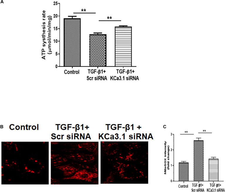
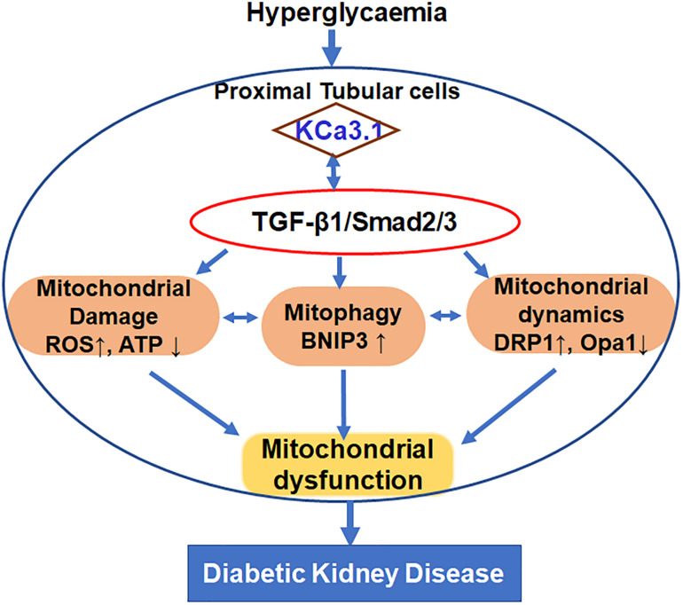
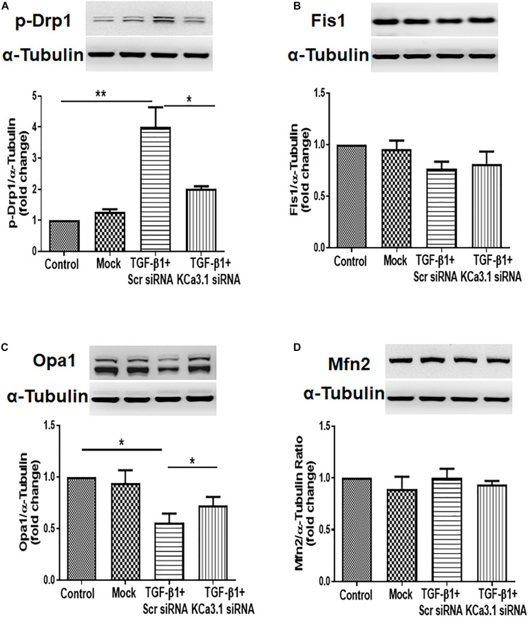
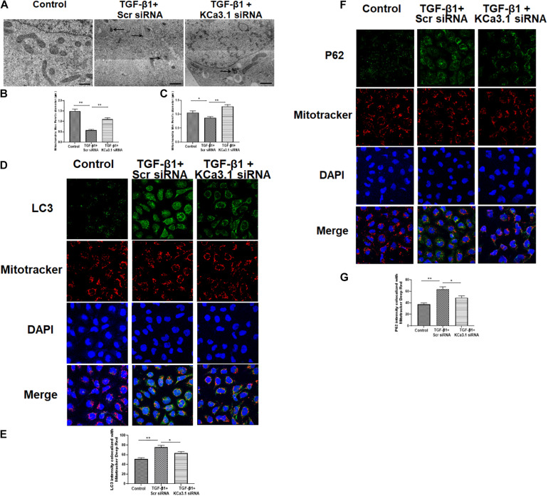
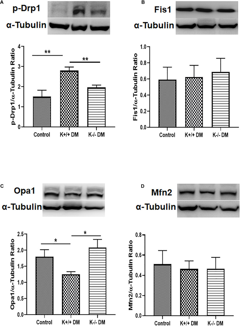
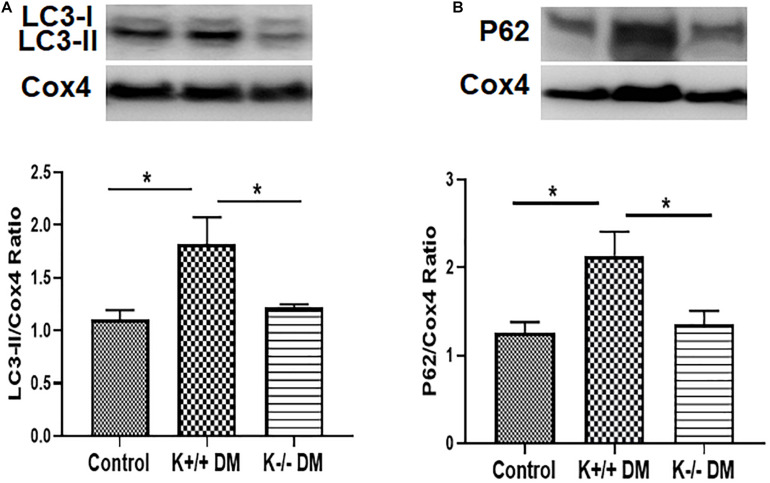
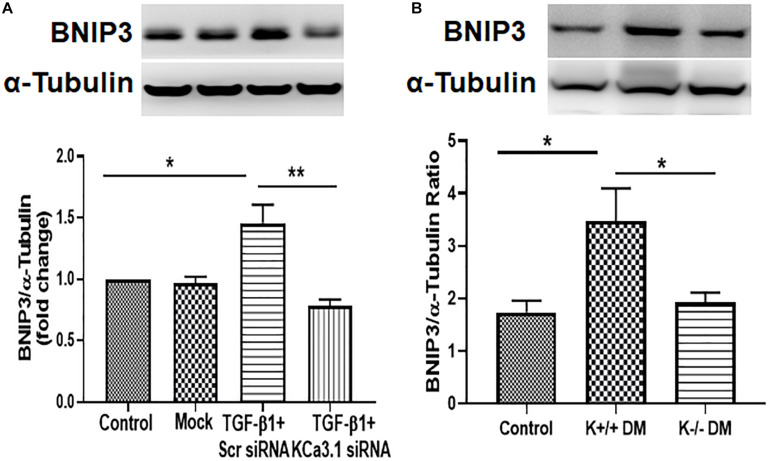
Mitochondrial dysfunction is implicated in the pathogenesis of diabetic kidney disease. Mitochondrial quality control is primarily mediated by mitochondrial turnover and repair through mitochondrial fission/fusion and mitophagy. We have previously shown that blockade of the calcium-activated potassium channel KCa3.1 ameliorates diabetic renal fibrosis. However, the mechanistic link between KCa3.1 and mitochondrial quality control in diabetic kidney disease is not yet known. Transforming growth factor β1 (TGF-β1) plays a central role in diabetic kidney disease. Recent studies indicate an emerging role of TGF-β1 in the regulation of mitochondrial function. However, the molecular mechanism mediating mitochondrial quality control in response to TGF-β1 remains limited. In this study, mitochondrial function was assessed in TGF-β1-exposed renal proximal tubular epithelial cells (HK2 cells) transfected with scrambled siRNA or KCa3.1 siRNA. , diabetes was induced in KCa3.1+/+ and KCa3.1-/- mice by low-dose streptozotocin (STZ) injection. Mitochondrial fission/fusion-related proteins and mitophagy markers, as well as BCL2 interacting protein 3 (BNIP3) (a mitophagy regulator) were examined in HK2 cells and diabetic mice kidneys. The results showed that TGF-β1 significantly inhibited mitochondrial ATP production rate and increased mitochondrial ROS (mtROS) production when compared to control, which was normalized by KCa3.1 gene silencing. Increased fission and suppressed fusion were found in both TGF-β1-treated HK2 cells and diabetic mice, which were reversed by KCa3.1 deficiency. Furthermore, our results showed that mitophagy was inhibited in both and models of diabetic kidney disease. KCa3.1 deficiency restored abnormal mitophagy by inhibiting BNIP3 expression in TGF-β1-induced HK2 cells as well as in the diabetic mice. Collectively, these results indicate that KCa3.1 mediates the dysregulation of mitochondrial quality control in diabetic kidney disease.

摘要

线粒体功能障碍与糖尿病肾病的发病机制有关。线粒体质量控制主要通过线粒体分裂/融合和线粒体自噬介导的线粒体更新和修复来实现。我们之前已经表明,钙激活钾通道KCa3.1的阻断可改善糖尿病肾纤维化。然而,在糖尿病肾病中,KCa3.1与线粒体质量控制之间的机制联系尚不清楚。转化生长因子β1(TGF-β1)在糖尿病肾病中起核心作用。最近的研究表明,TGF-β1在调节线粒体功能方面有新的作用。然而,介导对TGF-β1反应的线粒体质量控制的分子机制仍然有限。在本研究中,在用乱序siRNA或KCa3.1 siRNA转染的TGF-β1处理的肾近端小管上皮细胞(HK2细胞)中评估线粒体功能。通过低剂量链脲佐菌素(STZ)注射在KCa3.1+/+和KCa3.1-/-小鼠中诱导糖尿病。在HK2细胞和糖尿病小鼠肾脏中检测线粒体分裂/融合相关蛋白和线粒体自噬标志物,以及BCL2相互作用蛋白3(BNIP3)(一种线粒体自噬调节因子)。结果表明,与对照相比,TGF-β1显著抑制线粒体ATP产生率并增加线粒体ROS(mtROS)产生,而KCa3.1基因沉默可使其恢复正常。在TGF-β1处理的HK2细胞和糖尿病小鼠中均发现裂变增加和融合受抑制,而KCa3.1缺乏可使其逆转。此外,我们的结果表明,在糖尿病肾病的两种模型中,线粒体自噬均受到抑制。KCa3.1缺乏通过抑制TGF-β1诱导的HK2细胞以及糖尿病小鼠中的BNIP3表达来恢复异常的线粒体自噬。总的来说,这些结果表明KCa3.1介导糖尿病肾病中线粒体质量控制的失调。

https://cdn.ncbi.nlm.nih.gov/pmc/blobs/7c48/7933228/4f8ed23ff9a8/fcell-09-573814-g007.jpg
https://cdn.ncbi.nlm.nih.gov/pmc/blobs/7c48/7933228/9c9bbf64520a/fcell-09-573814-g001.jpg
https://cdn.ncbi.nlm.nih.gov/pmc/blobs/7c48/7933228/0e4648090cee/fcell-09-573814-g002.jpg
https://cdn.ncbi.nlm.nih.gov/pmc/blobs/7c48/7933228/8d26fe3cce0a/fcell-09-573814-g003.jpg
https://cdn.ncbi.nlm.nih.gov/pmc/blobs/7c48/7933228/5b428022302d/fcell-09-573814-g004.jpg
https://cdn.ncbi.nlm.nih.gov/pmc/blobs/7c48/7933228/00e753708716/fcell-09-573814-g005.jpg
https://cdn.ncbi.nlm.nih.gov/pmc/blobs/7c48/7933228/01e822fc3d6c/fcell-09-573814-g006.jpg
https://cdn.ncbi.nlm.nih.gov/pmc/blobs/7c48/7933228/4f8ed23ff9a8/fcell-09-573814-g007.jpg

相似文献

1
KCa3.1 Mediates Dysregulation of Mitochondrial Quality Control in Diabetic Kidney Disease.KCa3.1介导糖尿病肾病中线粒体质量控制的失调。
Front Cell Dev Biol. 2021 Feb 19;9:573814. doi: 10.3389/fcell.2021.573814. eCollection 2021.
2
KCa3.1 mediates dysfunction of tubular autophagy in diabetic kidneys via PI3k/Akt/mTOR signaling pathways.KCa3.1 通过 PI3k/Akt/mTOR 信号通路介导糖尿病肾脏管状自噬功能障碍。
Sci Rep. 2016 Mar 31;6:23884. doi: 10.1038/srep23884.
3
Inhibition of KCa3.1 suppresses TGF-β1 induced MCP-1 expression in human proximal tubular cells through Smad3, p38 and ERK1/2 signaling pathways.抑制KCa3.1可通过Smad3、p38和ERK1/2信号通路抑制转化生长因子-β1诱导的人近端肾小管细胞中单核细胞趋化蛋白-1的表达。
Int J Biochem Cell Biol. 2014 Feb;47:1-10. doi: 10.1016/j.biocel.2013.11.017. Epub 2013 Nov 28.
4
Blockade of KCa3.1 ameliorates renal fibrosis through the TGF-β1/Smad pathway in diabetic mice.阻断 KCa3.1 通过 TGF-β1/Smad 通路减轻糖尿病小鼠的肾纤维化。
Diabetes. 2013 Aug;62(8):2923-34. doi: 10.2337/db13-0135. Epub 2013 May 8.
5
High glucose induces CCL20 in proximal tubular cells via activation of the KCa3.1 channel.高糖通过激活KCa3.1通道诱导近端肾小管细胞产生CCL20。
PLoS One. 2014 Apr 14;9(4):e95173. doi: 10.1371/journal.pone.0095173. eCollection 2014.
6
KCa3.1 mediates activation of fibroblasts in diabetic renal interstitial fibrosis.KCa3.1介导糖尿病肾间质纤维化中纤维母细胞的激活。
Nephrol Dial Transplant. 2014 Feb;29(2):313-24. doi: 10.1093/ndt/gft431. Epub 2013 Oct 28.
7
Huangkui capsule attenuates diabetic kidney disease through the induction of mitophagy mediated by STING1/PINK1 signaling in tubular cells.黄葵胶囊通过 STING1/PINK1 信号介导的小管细胞自噬来减轻糖尿病肾病。
Phytomedicine. 2023 Oct;119:154975. doi: 10.1016/j.phymed.2023.154975. Epub 2023 Jul 18.
8
Acetylshikonin from Zicao ameliorates renal dysfunction and fibrosis in diabetic mice by inhibiting TGF-β1/Smad pathway.紫草素通过抑制 TGF-β1/Smad 通路改善糖尿病小鼠的肾功能障碍和纤维化。
Hum Cell. 2018 Jul;31(3):199-209. doi: 10.1007/s13577-017-0192-8. Epub 2018 Mar 17.
9
Thioredoxin interacting protein (TXNIP) regulates tubular autophagy and mitophagy in diabetic nephropathy through the mTOR signaling pathway.硫氧还蛋白相互作用蛋白 (TXNIP) 通过 mTOR 信号通路调节糖尿病肾病中的管状自噬和线粒体自噬。
Sci Rep. 2016 Jul 6;6:29196. doi: 10.1038/srep29196.
10
KCa3.1 in diabetic kidney disease.糖尿病肾病中的KCa3.1
Curr Opin Nephrol Hypertens. 2022 Jan 1;31(1):129-134. doi: 10.1097/MNH.0000000000000751.

引用本文的文献

1
Mitochondrial quality control in diabetes mellitus and complications: molecular mechanisms and therapeutic strategies.糖尿病及其并发症中的线粒体质量控制:分子机制与治疗策略
Cell Death Dis. 2025 Aug 27;16(1):652. doi: 10.1038/s41419-025-07936-y.
2
Important regulatory role of mitophagy in diabetic microvascular complications.线粒体自噬在糖尿病微血管并发症中的重要调节作用。
J Transl Med. 2025 Mar 4;23(1):269. doi: 10.1186/s12967-025-06307-7.
3
Omega-3 Fatty Acids Modify Drp1 Expression and Activate the PINK1-Dependent Mitophagy Pathway in the Kidney and Heart of Adenine-Induced Uremic Rats.

本文引用的文献

1
Sitagliptin ameliorates renal tubular injury in diabetic kidney disease via STAT3-dependent mitochondrial homeostasis through SDF-1α/CXCR4 pathway.西他列汀通过 SDF-1α/CXCR4 通路改善糖尿病肾病肾小管损伤,通过 STAT3 依赖的线粒体稳态。
FASEB J. 2020 Jun;34(6):7500-7519. doi: 10.1096/fj.201903038R. Epub 2020 Apr 12.
2
Huangqi-Danshen decoction alleviates diabetic nephropathy in mice by inhibiting PINK1/Parkin-mediated mitophagy.黄芪丹参汤通过抑制PINK1/Parkin介导的线粒体自噬减轻小鼠糖尿病肾病。
Am J Transl Res. 2020 Mar 15;12(3):989-998. eCollection 2020.
3
Increased mitochondrial fragmentation in polycystic kidney disease acts as a modifier of disease progression.
ω-3脂肪酸调节腺嘌呤诱导的尿毒症大鼠肾脏和心脏中Drp1的表达并激活依赖PINK1的线粒体自噬途径。
Biomedicines. 2024 Sep 15;12(9):2107. doi: 10.3390/biomedicines12092107.
4
Integrated oral microgel system ameliorates renal fibrosis by hitchhiking co-delivery and targeted gut flora modulation.整合口腔微凝胶系统通过搭乘共递送和靶向肠道菌群调节改善肾纤维化。
J Nanobiotechnology. 2024 Jun 1;22(1):305. doi: 10.1186/s12951-024-02586-2.
5
The key mediator of diabetic kidney disease: Potassium channel dysfunction.糖尿病肾病的关键介质:钾通道功能障碍。
Genes Dis. 2023 Sep 22;11(4):101119. doi: 10.1016/j.gendis.2023.101119. eCollection 2024 Jul.
6
Dysfunctional Mitochondria Clearance in Situ: Mitophagy in Obesity and Diabetes-Associated Cardiometabolic Diseases.功能失调的线粒体原位清除:肥胖和糖尿病相关代谢性心血管疾病中的自噬。
Diabetes Metab J. 2024 Jul;48(4):503-517. doi: 10.4093/dmj.2023.0213. Epub 2024 Feb 15.
7
Effects of honokiol protects against chronic kidney disease via BNIP3/NIX and FUNDC1-mediated mitophagy and AMPK pathways.和厚朴酚通过 BNIP3/NIX 和 FUNDC1 介导的线粒体自噬和 AMPK 通路对慢性肾脏病的保护作用。
Mol Biol Rep. 2023 Aug;50(8):6557-6568. doi: 10.1007/s11033-023-08592-1. Epub 2023 Jun 20.
8
BET Protein Inhibitor JQ1 Modulates Mitochondrial Dysfunction and Oxidative Stress Induced by Chronic Kidney Disease.BET蛋白抑制剂JQ1调节慢性肾脏病诱导的线粒体功能障碍和氧化应激。
Antioxidants (Basel). 2023 May 20;12(5):1130. doi: 10.3390/antiox12051130.
9
ATF5 regulates tubulointerstitial injury in diabetic kidney disease via mitochondrial unfolded protein response.ATF5 通过线粒体未折叠蛋白反应调节糖尿病肾病的肾小管间质损伤。
Mol Med. 2023 Apr 24;29(1):57. doi: 10.1186/s10020-023-00651-4.
10
KCa3.1 Promotes Proinflammatory Exosome Secretion by Activating AKT/Rab27a in Atrial Myocytes during Rapid Pacing.快速起搏时 KCa3.1 通过激活心房肌细胞中的 AKT/Rab27a 促进促炎小体分泌。
Cardiovasc Ther. 2023 Mar 30;2023:3939360. doi: 10.1155/2023/3939360. eCollection 2023.
多囊肾病中线粒体碎片化增加可作为疾病进展的修饰因子。
FASEB J. 2020 May;34(5):6493-6507. doi: 10.1096/fj.201901739RR. Epub 2020 Apr 2.
4
Bnip3 in mitophagy: Novel insights and potential therapeutic target for diseases of secondary mitochondrial dysfunction.Bnip3 在自噬中的作用:继发性线粒体功能障碍疾病的新见解和潜在治疗靶点。
Clin Chim Acta. 2020 Jul;506:72-83. doi: 10.1016/j.cca.2020.02.024. Epub 2020 Feb 21.
5
Activation of TRPV1 channel antagonizes diabetic nephropathy through inhibiting endoplasmic reticulum-mitochondria contact in podocytes.TRPV1 通道的激活通过抑制足细胞内质网-线粒体接触来拮抗糖尿病肾病。
Metabolism. 2020 Apr;105:154182. doi: 10.1016/j.metabol.2020.154182. Epub 2020 Feb 13.
6
Drp1-mediated mitochondrial fission promotes renal fibroblast activation and fibrogenesis.Drp1 介导线粒体分裂促进肾成纤维细胞活化和纤维化。
Cell Death Dis. 2020 Jan 16;11(1):29. doi: 10.1038/s41419-019-2218-5.
7
Fundc1-dependent mitophagy is obligatory to ischemic preconditioning-conferred renoprotection in ischemic AKI via suppression of Drp1-mediated mitochondrial fission.Fundc1 依赖性线粒体自噬对于缺血预处理诱导的肾保护是必需的,其通过抑制 Drp1 介导的线粒体分裂来实现。
Redox Biol. 2020 Feb;30:101415. doi: 10.1016/j.redox.2019.101415. Epub 2019 Dec 28.
8
Blockage of ROS-ERK-DLP1 signaling and mitochondrial fission alleviates Cr(VI)-induced mitochondrial dysfunction in L02 hepatocytes.阻断 ROS-ERK-DLP1 信号通路和线粒体分裂可减轻 Cr(VI)诱导的 L02 肝细胞线粒体功能障碍。
Ecotoxicol Environ Saf. 2019 Dec 30;186:109749. doi: 10.1016/j.ecoenv.2019.109749. Epub 2019 Oct 14.
9
PGC-1α Suppresses the Activation of TGF-β/Smad Signaling via Targeting TGFβRI Downregulation by Upregulation.PGC-1α 通过上调 TGFβRI 的表达来抑制 TGF-β/Smad 信号通路的激活。
Int J Mol Sci. 2019 Oct 14;20(20):5084. doi: 10.3390/ijms20205084.
10
Activation of BNIP3-mediated mitophagy protects against renal ischemia-reperfusion injury.BNIP3 介导线粒体自噬对肾缺血再灌注损伤起保护作用。
Cell Death Dis. 2019 Sep 12;10(9):677. doi: 10.1038/s41419-019-1899-0.