• 文献检索
  • 文档翻译
  • 深度研究
  • 学术资讯
  • Suppr Zotero 插件Zotero 插件
  • 邀请有礼
  • 套餐&价格
  • 历史记录
应用&插件
Suppr Zotero 插件Zotero 插件浏览器插件Mac 客户端Windows 客户端微信小程序
定价
高级版会员购买积分包购买API积分包
服务
文献检索文档翻译深度研究API 文档MCP 服务
关于我们
关于 Suppr公司介绍联系我们用户协议隐私条款
关注我们

Suppr 超能文献

核心技术专利:CN118964589B侵权必究
粤ICP备2023148730 号-1Suppr @ 2026

文献检索

告别复杂PubMed语法,用中文像聊天一样搜索,搜遍4000万医学文献。AI智能推荐,让科研检索更轻松。

立即免费搜索

文件翻译

保留排版,准确专业,支持PDF/Word/PPT等文件格式,支持 12+语言互译。

免费翻译文档

深度研究

AI帮你快速写综述,25分钟生成高质量综述,智能提取关键信息,辅助科研写作。

立即免费体验

新型冠状病毒肺炎的神经及神经影像学表现谱

Spectrum of neurologic & neuroimaging manifestation in COVID-19.

作者信息

Rehmani Razia, Segan Scott, Maddika Srikanth Reddy, Lei Yadanar Win, Broka Andrea

机构信息

Chief of Neuroradiology & Musculoskeletal Imaging, Department of Radiology, SBH Health System, NY, USA.

Chief of Neurology SBH Health System, Department of Neurology, SBH Health System, NY, USA.

出版信息

Brain Behav Immun Health. 2021 May;13:100238. doi: 10.1016/j.bbih.2021.100238. Epub 2021 Mar 3.

DOI:10.1016/j.bbih.2021.100238
PMID:33681827
原文链接:https://pmc.ncbi.nlm.nih.gov/articles/PMC7925234/
Abstract

OBJECTIVE

Coronavirus Disease 2019 (COVID-19) initially thought to be confined to the respiratory system only, is now known to be a multisystem disease. It is critical to be aware of and timely recognize neurological and neuroradiological manifestations affecting patients with COVID-19, to minimize morbidity and mortality of affected patients.

METHODS

We performed a retrospective chart review of patients admitted to our Level 1 trauma and stroke center during the peak of the COVID-19 outbreak in New York from March 1st to May 30, 2020, with a positive test for Severe Acute Respiratory Syndrome Coronavirus 2 (SARS-CoV-2) who presented mainly with neurological findings and had acute radiological brain changes on Computed Tomography (CT) scan. Patients with known chronic neurological disease processes were excluded from the study. We obtained and reviewed demographics, complete blood count, metabolic panel, D-dimer, inflammatory markers such as erythrocyte sedimentation rate (ESR), C reactive protein (CRP), imaging, and patient's hospital course. We reviewed the current literature on neuroimaging, pathophysiology, and their clinical correlations on COVID-19. This case series study was approved by our institutional review board.

RESULT

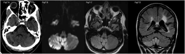
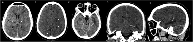
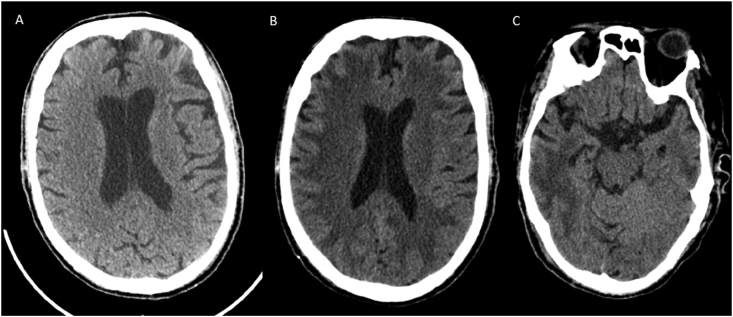
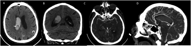
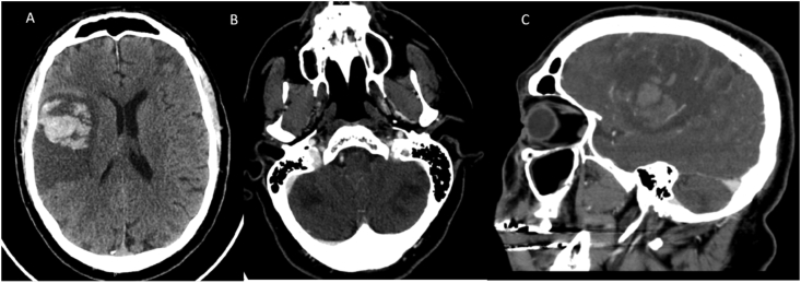
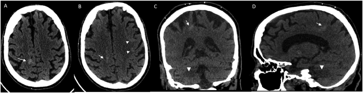
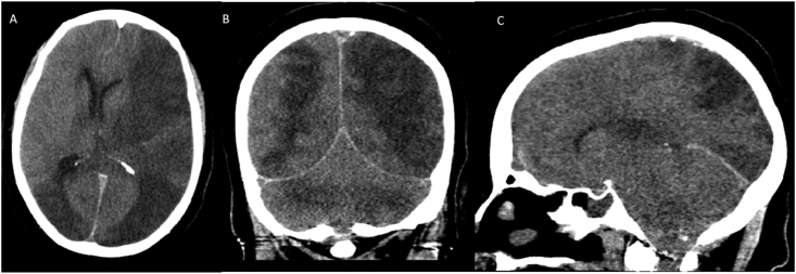
A total of 16 patients were selected for our case series. The most common neuroimaging features on CT, were territorial to multifocal ischemic infarcts, followed by a combination of ischemia and acute white matter encephalopathic changes, followed by temporal lobe predominant focal or more generalized encephalopathy with both confluent and non-confluent patterns, isolated cortical or more extensive intracranial hemorrhages and some combination of ischemia or hemorrhage and white matter changes. All our patients had severe acute respiratory distress syndrome (ARDS), most of them had elevated inflammatory markers, and D dimer.

CONCLUSION

COVID-19 infection is a multi-organ disease, which can manifest as rapidly progressive neurological disease beyond the more common pulmonary presentation. Early recognition of various neurological findings and neuroimaging patterns in these patients will enable timely diagnosis and rapid treatment to reduce morbidity and mortality. Our retrospective study is limited due to small non-representative sample size, strict selection criteria likely underestimating the true extent of neurological manifestations of COVID-19, mono-modality imaging technique limited to predominantly CT scans and lack of CSF analysis in all except one patient. Multi-institutional, multi-modality, largescale studies are needed with radio-pathological correlation to better understand the complete spectrum of neurologic presentations in COVID-19 patients and study the causal relationship between SARS-CoV-2 and CNS disease process.

https://cdn.ncbi.nlm.nih.gov/pmc/blobs/6f0b/8474170/78dc03919ed2/gr7.jpg
https://cdn.ncbi.nlm.nih.gov/pmc/blobs/6f0b/8474170/8622a1480ba7/gr1.jpg
https://cdn.ncbi.nlm.nih.gov/pmc/blobs/6f0b/8474170/a9d921c4b08e/gr2.jpg
https://cdn.ncbi.nlm.nih.gov/pmc/blobs/6f0b/8474170/ea352a97ebae/gr3.jpg
https://cdn.ncbi.nlm.nih.gov/pmc/blobs/6f0b/8474170/0e1d5d606deb/gr4.jpg
https://cdn.ncbi.nlm.nih.gov/pmc/blobs/6f0b/8474170/36cfefe24b93/gr5.jpg
https://cdn.ncbi.nlm.nih.gov/pmc/blobs/6f0b/8474170/7d5df9fc9e92/gr6.jpg
https://cdn.ncbi.nlm.nih.gov/pmc/blobs/6f0b/8474170/78dc03919ed2/gr7.jpg
摘要

目的

2019冠状病毒病(COVID-19)最初被认为仅局限于呼吸系统,现在已知是一种多系统疾病。了解并及时识别影响COVID-19患者的神经和神经放射学表现,对于将受影响患者的发病率和死亡率降至最低至关重要。

方法

我们对2020年3月1日至5月30日纽约COVID-19疫情高峰期入住我们一级创伤和卒中中心的患者进行了回顾性病历审查,这些患者严重急性呼吸综合征冠状病毒2(SARS-CoV-2)检测呈阳性,主要表现为神经学症状,且计算机断层扫描(CT)显示脑部有急性放射学改变。已知患有慢性神经疾病的患者被排除在研究之外。我们获取并审查了人口统计学数据、全血细胞计数、代谢指标、D-二聚体、炎症标志物如红细胞沉降率(ESR)、C反应蛋白(CRP)、影像学检查以及患者的住院病程。我们回顾了当前关于COVID-19神经影像学、病理生理学及其临床相关性的文献。本病例系列研究已获得我们机构审查委员会的批准。

结果

我们的病例系列共纳入16例患者。CT上最常见的神经影像学特征是从区域性到多灶性缺血性梗死,其次是缺血与急性白质脑病改变并存,然后是以颞叶为主的局灶性或更广泛的脑病,有融合和非融合模式,孤立的皮质或更广泛的颅内出血,以及缺血或出血与白质改变的某种组合。我们所有患者均患有严重急性呼吸窘迫综合征(ARDS),大多数患者炎症标志物和D-二聚体升高。

结论

COVID-19感染是一种多器官疾病,除了更常见的肺部表现外,还可表现为快速进展的神经疾病。早期识别这些患者的各种神经学表现和神经影像学模式将有助于及时诊断和快速治疗,以降低发病率和死亡率。我们的回顾性研究存在局限性,因为样本量小且不具代表性,严格的选择标准可能低估了COVID-19神经学表现的真实程度,单一的影像学技术主要限于CT扫描,且除一名患者外所有患者均未进行脑脊液分析。需要开展多机构、多模式、大规模研究,并进行放射病理学相关性分析,以更好地了解COVID-19患者神经学表现的全貌,并研究SARS-CoV-2与中枢神经系统疾病过程之间的因果关系。

相似文献

1
Spectrum of neurologic & neuroimaging manifestation in COVID-19.新型冠状病毒肺炎的神经及神经影像学表现谱
Brain Behav Immun Health. 2021 May;13:100238. doi: 10.1016/j.bbih.2021.100238. Epub 2021 Mar 3.
2
Neuroimaging manifestations in children with SARS-CoV-2 infection: a multinational, multicentre collaborative study.儿童严重急性呼吸综合征冠状病毒 2 感染的神经影像学表现:一项多国家、多中心合作研究。
Lancet Child Adolesc Health. 2021 Mar;5(3):167-177. doi: 10.1016/S2352-4642(20)30362-X. Epub 2020 Dec 16.
3
A Retrospective Observational Study of Neurological Manifestations in COVID-19 (SON-CoV).一项关于 COVID-19(SON-CoV)神经系统表现的回顾性观察研究。
J Assoc Physicians India. 2022 Oct;70(10):11-12. doi: 10.5005/japi-11001-0107.
4
Pediatrics COVID-19 and neurological manifestations: Single tertiary centre experience.儿科新型冠状病毒肺炎与神经表现:单一三级医疗中心的经验
eNeurologicalSci. 2021 Sep;24:100355. doi: 10.1016/j.ensci.2021.100355. Epub 2021 Jul 9.
5
Human and novel coronavirus infections in children: a review.儿童人感染和新型冠状病毒感染:综述。
Paediatr Int Child Health. 2021 Feb;41(1):36-55. doi: 10.1080/20469047.2020.1781356. Epub 2020 Jun 25.
6
Correlation of neuroimaging and thorax CT findings in patients with COVID-19: A large single-center experience.新型冠状病毒肺炎患者神经影像学与胸部 CT 表现的相关性:一项大型单中心经验。
Turk J Med Sci. 2021 Dec 13;51(6):2850-2860. doi: 10.3906/sag-2105-138.
7
COVID-19-related intracranial imaging findings: a large single-centre experience.COVID-19 相关的颅内影像学表现:一项大型单中心研究。
Clin Radiol. 2021 Feb;76(2):108-116. doi: 10.1016/j.crad.2020.09.002. Epub 2020 Sep 15.
8
Neuroimaging findings and neurological manifestations in hospitalized COVID-19 patients: Impact of cancer and ventilatory support status.住院 COVID-19 患者的神经影像学表现和神经系统表现:癌症和通气支持状态的影响。
PLoS One. 2023 Mar 24;18(3):e0283614. doi: 10.1371/journal.pone.0283614. eCollection 2023.
9
Neurological symptoms in COVID-19: a cross-sectional monocentric study of hospitalized patients.新型冠状病毒肺炎的神经系统症状:一项针对住院患者的横断面单中心研究
Neurol Res Pract. 2021 Mar 12;3(1):17. doi: 10.1186/s42466-021-00116-1.
10
Abnormalities of brain imaging in COVID-19 patients with neurological symptoms.患有神经系统症状的新冠病毒肺炎患者的脑成像异常
Curr J Neurol. 2023 Jul 6;22(3):162-169. doi: 10.18502/cjn.v22i3.13796.

引用本文的文献

1
Presumed Fourth Nerve Palsy in a Healthy and Asymptomatic Child with COVID-19 Infection.一名感染新冠病毒的健康无症状儿童疑似动眼神经麻痹。
Iran J Child Neurol. 2024 Summer;18(3):137-141. doi: 10.22037/v18i3.42705. Epub 2024 Jun 22.
2
Impact of Symptomatology, Clinical and Radiological Severity of COVID-19 on Pulmonary Function Test Results and Functional Capacity during Follow-Up among Survivors.COVID-19的症状学、临床及影像学严重程度对幸存者随访期间肺功能测试结果及功能能力的影响
J Clin Med. 2023 Dec 21;13(1):45. doi: 10.3390/jcm13010045.
3
Topographical Distribution of Neuroanatomical Abnormalities Following COVID-19 Invasion : A Systematic Literature Review.

本文引用的文献

1
ACE2: Evidence of role as entry receptor for SARS-CoV-2 and implications in comorbidities.ACE2:作为 SARS-CoV-2 进入受体的证据及其在合并症中的意义。
Elife. 2020 Nov 9;9:e61390. doi: 10.7554/eLife.61390.
2
Intracranial hemorrhage in coronavirus disease 2019 (COVID-19) patients.COVID-19 患者的颅内出血。
Neurol Sci. 2021 Jan;42(1):25-33. doi: 10.1007/s10072-020-04870-z. Epub 2020 Nov 3.
3
Guillain-Barré syndrome in SARS-CoV-2 infection: an instant systematic review of the first six months of pandemic.严重急性呼吸综合征冠状病毒 2 感染相关格林-巴利综合征:大流行头 6 个月的即时系统综述。
新冠病毒入侵后神经解剖结构异常的分布:一项系统文献综述。
Clin Neuroradiol. 2024 Mar;34(1):13-31. doi: 10.1007/s00062-023-01344-5. Epub 2023 Sep 11.
4
Neurological manifestation in COVID-19 disease with neuroimaging studies.新型冠状病毒肺炎(COVID-19)疾病的神经学表现及神经影像学研究
Am J Neurodegener Dis. 2023 Apr 15;12(2):42-84. eCollection 2023.
5
Comparative imaging findings among different primary betacoronaviruses.不同原发性β冠状病毒之间的对比影像学表现。
Pol J Radiol. 2022 Nov 30;87:e641-e651. doi: 10.5114/pjr.2022.121763. eCollection 2022.
6
COVID-19: Invasion, pathogenesis and possible cure - A review.COVID-19:入侵、发病机制和可能的治疗方法 - 综述。
J Virol Methods. 2022 Feb;300:114434. doi: 10.1016/j.jviromet.2021.114434. Epub 2021 Dec 14.
7
[COVID-19: neurological manifestations-update : What we know so far].[新型冠状病毒肺炎:神经学表现——最新进展:目前我们所了解的情况]
Radiologe. 2021 Oct;61(10):902-908. doi: 10.1007/s00117-021-00907-2. Epub 2021 Sep 9.
8
Unilateral diplopia and ptosis in a child with COVID-19 revealing third cranial nerve palsy.儿童感染 COVID-19 后出现单侧复视和上睑下垂,提示动眼神经麻痹。
J Infect Public Health. 2021 Sep;14(9):1198-1200. doi: 10.1016/j.jiph.2021.08.007. Epub 2021 Aug 10.
J Neurol Neurosurg Psychiatry. 2020 Oct;91(10):1105-1110. doi: 10.1136/jnnp-2020-324491. Epub 2020 Aug 27.
4
The association of race and COVID-19 mortality.种族与新冠病毒疾病死亡率之间的关联。
EClinicalMedicine. 2020 Aug;25:100455. doi: 10.1016/j.eclinm.2020.100455. Epub 2020 Jul 15.
5
The emerging spectrum of COVID-19 neurology: clinical, radiological and laboratory findings.新型冠状病毒肺炎神经系统表现的研究进展:临床、影像学和实验室表现。
Brain. 2020 Oct 1;143(10):3104-3120. doi: 10.1093/brain/awaa240.
6
Neuroimaging and neurologic findings in COVID-19 and other coronavirus infections: A systematic review in 116 patients.COVID-19 及其他冠状病毒感染的神经影像学和神经学表现:116 例患者的系统评价。
J Neuroradiol. 2021 Feb;48(1):43-50. doi: 10.1016/j.neurad.2020.06.007. Epub 2020 Jun 27.
7
Early evidence of pronounced brain involvement in fatal COVID-19 outcomes.有明显脑部受累与COVID-19致命结局相关的早期证据。
Lancet. 2020 Jun 20;395(10241):e109. doi: 10.1016/S0140-6736(20)31282-4. Epub 2020 Jun 4.
8
Brain Imaging Use and Findings in COVID-19: A Single Academic Center Experience in the Epicenter of Disease in the United States.脑影像学在 COVID-19 中的应用及结果:美国疫情中心单一学术中心的经验。
AJNR Am J Neuroradiol. 2020 Jul;41(7):1179-1183. doi: 10.3174/ajnr.A6610. Epub 2020 May 28.
9
COVID-19-associated hyperviscosity: a link between inflammation and thrombophilia?新冠病毒感染相关的高黏滞血症:炎症与血栓形成倾向之间的联系?
Lancet. 2020 Jun 6;395(10239):1758-1759. doi: 10.1016/S0140-6736(20)31209-5. Epub 2020 May 25.
10
Spontaneous echo contrast in venous ultrasound of severe COVID-19 patients.重症 COVID-19 患者静脉超声中的自发性回声造影
Intensive Care Med. 2020 Aug;46(8):1637-1639. doi: 10.1007/s00134-020-06094-3. Epub 2020 May 27.