• 文献检索
  • 文档翻译
  • 深度研究
  • 学术资讯
  • Suppr Zotero 插件Zotero 插件
  • 邀请有礼
  • 套餐&价格
  • 历史记录
应用&插件
Suppr Zotero 插件Zotero 插件浏览器插件Mac 客户端Windows 客户端微信小程序
定价
高级版会员购买积分包购买API积分包
服务
文献检索文档翻译深度研究API 文档MCP 服务
关于我们
关于 Suppr公司介绍联系我们用户协议隐私条款
关注我们

Suppr 超能文献

核心技术专利:CN118964589B侵权必究
粤ICP备2023148730 号-1Suppr @ 2026

文献检索

告别复杂PubMed语法,用中文像聊天一样搜索,搜遍4000万医学文献。AI智能推荐,让科研检索更轻松。

立即免费搜索

文件翻译

保留排版,准确专业,支持PDF/Word/PPT等文件格式,支持 12+语言互译。

免费翻译文档

深度研究

AI帮你快速写综述,25分钟生成高质量综述,智能提取关键信息,辅助科研写作。

立即免费体验

T 细胞受体信号强度对急性和慢性病毒感染时 CD4 T 细胞应答的相反影响。

Opposing effects of T cell receptor signal strength on CD4 T cells responding to acute versus chronic viral infection.

机构信息

Immune Cell Biology Laboratory, Department of Biomedicine, University of Basel, University Hospital Basel, Basel, Switzerland.

Division of Experimental Virology, Department of Biomedicine - Haus Petersplatz, University of Basel, Basel, Switzerland.

出版信息

Elife. 2021 Mar 8;10:e61869. doi: 10.7554/eLife.61869.

DOI:10.7554/eLife.61869
PMID:33684030
原文链接:https://pmc.ncbi.nlm.nih.gov/articles/PMC7943189/
Abstract

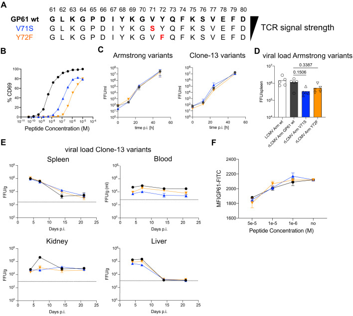
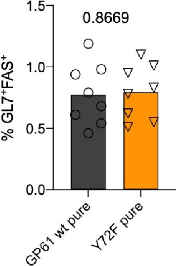
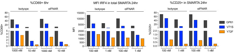
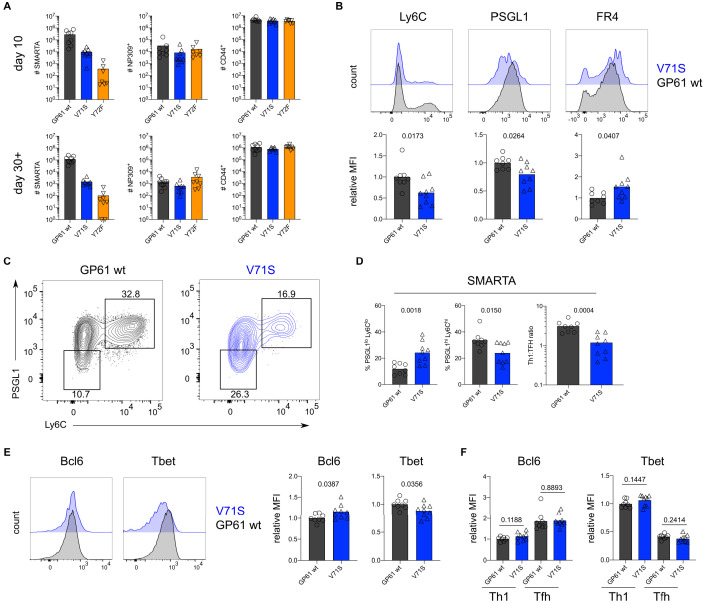
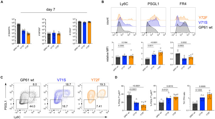
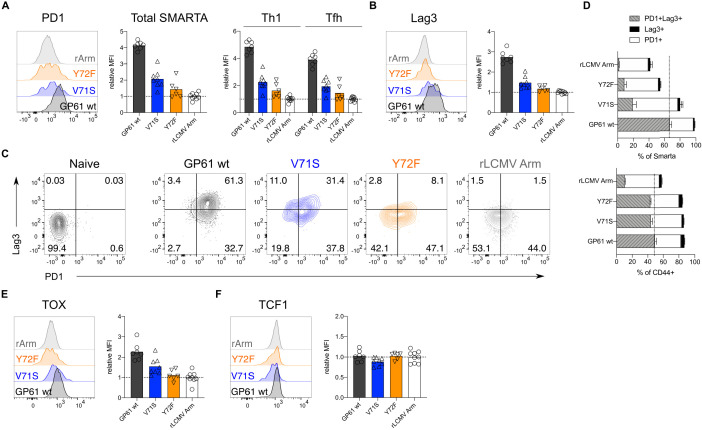
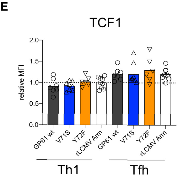
A hallmark of adaptive immunity is CD4 T cells' ability to differentiate into specialized effectors. A long-standing question is whether T cell receptor (TCR) signal strength can dominantly instruct the development of Th1 and T follicular helper (Tfh) cells across distinct infectious contexts. We characterized the differentiation of murine CD4 TCR transgenic T cells responding to altered peptide ligand lymphocytic choriomeningitis viruses (LCMV) derived from acute and chronic parental strains. We found that TCR signal strength exerts opposite and hierarchical effects on the balance of Th1 and Tfh cells responding to acute versus persistent infection. TCR signal strength correlates positively with Th1 generation during acute but negatively during chronic infection. Weakly activated T cells express lower levels of markers associated with chronic T cell stimulation and may resist functional inactivation. We anticipate that the panel of recombinant viruses described herein will be valuable for investigating a wide range of CD4 T cell responses.

摘要

适应性免疫的一个标志是 CD4 T 细胞将分化为专门的效应细胞的能力。一个长期存在的问题是,T 细胞受体(TCR)信号强度是否可以主导 Th1 和滤泡辅助 T 细胞(Tfh)细胞在不同感染环境下的发育。我们描述了对源自急性和慢性亲本株的改变肽配基淋巴细胞性脉络丛脑膜炎病毒(LCMV)的小鼠 CD4 TCR 转基因 T 细胞的分化。我们发现,TCR 信号强度对急性和持续性感染中 Th1 和 Tfh 细胞平衡的影响相反且具有层次结构。在急性感染期间,TCR 信号强度与 Th1 生成呈正相关,但在慢性感染期间呈负相关。弱激活的 T 细胞表达与慢性 T 细胞刺激相关的标志物水平较低,并且可能抵抗功能失活。我们预计本文描述的重组病毒将有助于研究广泛的 CD4 T 细胞反应。

https://cdn.ncbi.nlm.nih.gov/pmc/blobs/f40f/7943189/7a416f62d08d/elife-61869-resp-fig1.jpg
https://cdn.ncbi.nlm.nih.gov/pmc/blobs/f40f/7943189/02d1e76738e7/elife-61869-fig1.jpg
https://cdn.ncbi.nlm.nih.gov/pmc/blobs/f40f/7943189/bdfb5f8f3c04/elife-61869-fig1-figsupp1.jpg
https://cdn.ncbi.nlm.nih.gov/pmc/blobs/f40f/7943189/cc517f6fa763/elife-61869-fig1-figsupp2.jpg
https://cdn.ncbi.nlm.nih.gov/pmc/blobs/f40f/7943189/87c1f694539c/elife-61869-fig1-figsupp3.jpg
https://cdn.ncbi.nlm.nih.gov/pmc/blobs/f40f/7943189/d82669507374/elife-61869-fig2.jpg
https://cdn.ncbi.nlm.nih.gov/pmc/blobs/f40f/7943189/204b93f262d8/elife-61869-fig2-figsupp1.jpg
https://cdn.ncbi.nlm.nih.gov/pmc/blobs/f40f/7943189/e64b72e28527/elife-61869-fig2-figsupp2.jpg
https://cdn.ncbi.nlm.nih.gov/pmc/blobs/f40f/7943189/15bc50557380/elife-61869-fig2-figsupp3.jpg
https://cdn.ncbi.nlm.nih.gov/pmc/blobs/f40f/7943189/9b3ec2da2b74/elife-61869-fig2-figsupp4.jpg
https://cdn.ncbi.nlm.nih.gov/pmc/blobs/f40f/7943189/7644564174fe/elife-61869-fig2-figsupp5.jpg
https://cdn.ncbi.nlm.nih.gov/pmc/blobs/f40f/7943189/e61632342e2c/elife-61869-fig2-figsupp6.jpg
https://cdn.ncbi.nlm.nih.gov/pmc/blobs/f40f/7943189/0128207d2b9e/elife-61869-fig2-figsupp7.jpg
https://cdn.ncbi.nlm.nih.gov/pmc/blobs/f40f/7943189/804730e57bd4/elife-61869-fig3.jpg
https://cdn.ncbi.nlm.nih.gov/pmc/blobs/f40f/7943189/cb12b01bd96d/elife-61869-fig3-figsupp1.jpg
https://cdn.ncbi.nlm.nih.gov/pmc/blobs/f40f/7943189/03abe47c4e85/elife-61869-fig3-figsupp2.jpg
https://cdn.ncbi.nlm.nih.gov/pmc/blobs/f40f/7943189/c0e9eaa4a730/elife-61869-fig3-figsupp3.jpg
https://cdn.ncbi.nlm.nih.gov/pmc/blobs/f40f/7943189/4392dc01a597/elife-61869-fig3-figsupp4.jpg
https://cdn.ncbi.nlm.nih.gov/pmc/blobs/f40f/7943189/2333873836b4/elife-61869-fig3-figsupp5.jpg
https://cdn.ncbi.nlm.nih.gov/pmc/blobs/f40f/7943189/76a7c0422245/elife-61869-fig3-figsupp6.jpg
https://cdn.ncbi.nlm.nih.gov/pmc/blobs/f40f/7943189/72d7a2434bb4/elife-61869-fig3-figsupp7.jpg
https://cdn.ncbi.nlm.nih.gov/pmc/blobs/f40f/7943189/7100b37deca8/elife-61869-fig3-figsupp8.jpg
https://cdn.ncbi.nlm.nih.gov/pmc/blobs/f40f/7943189/1cb0e0a5da68/elife-61869-fig3-figsupp9.jpg
https://cdn.ncbi.nlm.nih.gov/pmc/blobs/f40f/7943189/9bb74bdedc48/elife-61869-fig3-figsupp10.jpg
https://cdn.ncbi.nlm.nih.gov/pmc/blobs/f40f/7943189/2dd628ad6481/elife-61869-fig4.jpg
https://cdn.ncbi.nlm.nih.gov/pmc/blobs/f40f/7943189/9d835bdbbed6/elife-61869-fig4-figsupp1.jpg
https://cdn.ncbi.nlm.nih.gov/pmc/blobs/f40f/7943189/f685325b4af5/elife-61869-fig4-figsupp2.jpg
https://cdn.ncbi.nlm.nih.gov/pmc/blobs/f40f/7943189/92e9e551c433/elife-61869-fig4-figsupp3.jpg
https://cdn.ncbi.nlm.nih.gov/pmc/blobs/f40f/7943189/b43c62dc2309/elife-61869-fig4-figsupp4.jpg
https://cdn.ncbi.nlm.nih.gov/pmc/blobs/f40f/7943189/3376107a96c9/elife-61869-fig4-figsupp5.jpg
https://cdn.ncbi.nlm.nih.gov/pmc/blobs/f40f/7943189/7a416f62d08d/elife-61869-resp-fig1.jpg

相似文献

1
Opposing effects of T cell receptor signal strength on CD4 T cells responding to acute versus chronic viral infection.T 细胞受体信号强度对急性和慢性病毒感染时 CD4 T 细胞应答的相反影响。
Elife. 2021 Mar 8;10:e61869. doi: 10.7554/eLife.61869.
2
Vaccine-elicited CD4 T cells prevent the deletion of antiviral B cells in chronic infection.疫苗诱导的 CD4 T 细胞可防止慢性感染中抗病毒 B 细胞的缺失。
Proc Natl Acad Sci U S A. 2021 Nov 16;118(46). doi: 10.1073/pnas.2108157118.
3
GITR intrinsically sustains early type 1 and late follicular helper CD4 T cell accumulation to control a chronic viral infection.糖皮质激素诱导肿瘤坏死因子受体(GITR)本质上维持早期1型和晚期滤泡辅助性CD4 T细胞的积累,以控制慢性病毒感染。
PLoS Pathog. 2015 Jan 15;11(1):e1004517. doi: 10.1371/journal.ppat.1004517. eCollection 2015 Jan.
4
PD-L1 Checkpoint Inhibition Narrows the Antigen-Specific T Cell Receptor Repertoire in Chronic Lymphocytic Choriomeningitis Virus Infection.PD-L1 检查点抑制缩小了慢性淋巴细胞脉络丛脑膜炎病毒感染中抗原特异性 T 细胞受体库。
J Virol. 2020 Aug 31;94(18). doi: 10.1128/JVI.00795-20.
5
T Cell Receptor Diversity and Lineage Relationship between Virus-Specific CD8 T Cell Subsets during Chronic Lymphocytic Choriomeningitis Virus Infection.慢性淋巴细胞脉络丛脑膜炎病毒感染过程中病毒特异性 CD8 T 细胞亚群间 T 细胞受体多样性和谱系关系。
J Virol. 2020 Sep 29;94(20). doi: 10.1128/JVI.00935-20.
6
Delineating the transcriptional landscape and clonal diversity of virus-specific CD4 T cells during chronic viral infection.描绘慢性病毒感染过程中病毒特异性 CD4 T 细胞的转录组图谱和克隆多样性。
Elife. 2022 Oct 18;11:e80079. doi: 10.7554/eLife.80079.
7
Virus-specific MHC-class II-restricted TCR-transgenic mice: effects on humoral and cellular immune responses after viral infection.病毒特异性MHC-II类限制性TCR转基因小鼠:病毒感染后对体液免疫和细胞免疫反应的影响。
Eur J Immunol. 1998 Jan;28(1):390-400. doi: 10.1002/(SICI)1521-4141(199801)28:01<390::AID-IMMU390>3.0.CO;2-O.
8
mTOR Promotes Antiviral Humoral Immunity by Differentially Regulating CD4 Helper T Cell and B Cell Responses.mTOR通过差异性调节CD4辅助性T细胞和B细胞反应来促进抗病毒体液免疫。
J Virol. 2017 Jan 31;91(4). doi: 10.1128/JVI.01653-16. Print 2017 Feb 15.
9
Pre-existing chromatin accessibility and gene expression differences among naive CD4 T cells influence effector potential.初始 CD4 T 细胞中预先存在的染色质可及性和基因表达差异影响效应潜力。
Cell Rep. 2021 Nov 30;37(9):110064. doi: 10.1016/j.celrep.2021.110064.
10
IRF9 Prevents CD8 T Cell Exhaustion in an Extrinsic Manner during Acute Lymphocytic Choriomeningitis Virus Infection.在急性淋巴细胞性脉络丛脑膜炎病毒感染期间,IRF9以非内在方式预防CD8 T细胞耗竭。
J Virol. 2017 Oct 27;91(22). doi: 10.1128/JVI.01219-17. Print 2017 Nov 15.

引用本文的文献

1
Engineering TCR-controlled fuzzy logic into CAR T cells enhances therapeutic specificity.将工程化TCR控制的模糊逻辑引入嵌合抗原受体T细胞可增强治疗特异性。
Cell. 2025 May 1;188(9):2372-2389.e35. doi: 10.1016/j.cell.2025.03.017. Epub 2025 Apr 11.
2
TRAF3 is critical for initial T follicular helper cell specification via coordination of the IL-6R/IL-2R-BCL6 signaling nexus.通过协调IL-6R/IL-2R-BCL6信号轴,TRAF3对于初始滤泡辅助性T细胞的分化至关重要。
Sci Immunol. 2025 Feb 14;10(104):eadr0517. doi: 10.1126/sciimmunol.adr0517.
3
CD6 regulates CD4 T follicular helper cell differentiation and humoral immunity during murine coronavirus infection.

本文引用的文献

1
TIGIT limits immune pathology during viral infections.TIGIT 限制病毒感染期间的免疫病理。
Nat Commun. 2020 Mar 9;11(1):1288. doi: 10.1038/s41467-020-15025-1.
2
Long-lived T follicular helper cells retain plasticity and help sustain humoral immunity.长寿的滤泡辅助性 T 细胞保持着可塑性,并有助于维持体液免疫。
Sci Immunol. 2020 Mar 6;5(45). doi: 10.1126/sciimmunol.aay5552.
3
The Transcription Factor Tox2 Drives T Follicular Helper Cell Development via Regulating Chromatin Accessibility.转录因子 Tox2 通过调节染色质可及性驱动滤泡辅助性 T 细胞的发育。
在小鼠冠状病毒感染期间,CD6调节CD4滤泡辅助性T细胞分化和体液免疫。
J Virol. 2025 Jan 31;99(1):e0186424. doi: 10.1128/jvi.01864-24. Epub 2024 Dec 16.
4
CD6 Regulates CD4 T Follicular Helper Cell Differentiation and Humoral Immunity During Murine Coronavirus Infection.CD6在小鼠冠状病毒感染期间调节CD4滤泡辅助性T细胞分化和体液免疫。
bioRxiv. 2024 Jul 26:2024.07.26.605237. doi: 10.1101/2024.07.26.605237.
5
Specialized Tfh cell subsets driving type-1 and type-2 humoral responses in lymphoid tissue.在淋巴组织中驱动1型和2型体液反应的特异性滤泡辅助性T细胞亚群。
Cell Discov. 2024 Jun 4;10(1):64. doi: 10.1038/s41421-024-00681-0.
6
Effector-Phase IL-2 Signals Drive Th1 Effector and Memory Responses Dependently and Independently of TCF-1.效应期 IL-2 信号依赖于 TCF-1 驱动 Th1 效应器和记忆应答 **解析**:“Effector-Phase”可直译为“效应期”,“IL-2”是“Interleukin-2”的缩写,可译为“白细胞介素-2”,“TCF-1”是“T-cell factor 1”的缩写,可译为“T 细胞因子 1”。
J Immunol. 2024 Feb 15;212(4):586-595. doi: 10.4049/jimmunol.2300570.
7
CD4 T cell memory.CD4 T 细胞记忆。
Nat Immunol. 2023 Jun;24(6):903-914. doi: 10.1038/s41590-023-01510-4. Epub 2023 May 8.
8
Toward a general model of CD4 T cell subset specification and memory cell formation.朝向 CD4 T 细胞亚群特化和记忆细胞形成的一般模型。
Immunity. 2023 Mar 14;56(3):475-484. doi: 10.1016/j.immuni.2023.02.010.
9
Delineating the transcriptional landscape and clonal diversity of virus-specific CD4 T cells during chronic viral infection.描绘慢性病毒感染过程中病毒特异性 CD4 T 细胞的转录组图谱和克隆多样性。
Elife. 2022 Oct 18;11:e80079. doi: 10.7554/eLife.80079.
10
Evolution of antigen-specific follicular helper T cell transcription from effector function to memory.从效应功能到记忆,抗原特异性滤泡辅助 T 细胞转录的演变。
Sci Immunol. 2022 Oct 14;7(76):eabm2084. doi: 10.1126/sciimmunol.abm2084. Epub 2022 Oct 7.
Immunity. 2019 Nov 19;51(5):826-839.e5. doi: 10.1016/j.immuni.2019.10.006. Epub 2019 Nov 12.
4
CD4 T cell exhaustion revealed by high PD-1 and LAG-3 expression and the loss of helper T cell function in chronic hepatitis B.慢性乙型肝炎中高表达 PD-1 和 LAG-3、辅助性 T 细胞功能丧失导致的 CD4 T 细胞耗竭。
BMC Immunol. 2019 Aug 7;20(1):27. doi: 10.1186/s12865-019-0309-9.
5
Single-cell RNA-seq reveals TOX as a key regulator of CD8 T cell persistence in chronic infection.单细胞 RNA 测序揭示 TOX 是慢性感染中 CD8 T 细胞持久性的关键调节因子。
Nat Immunol. 2019 Jul;20(7):890-901. doi: 10.1038/s41590-019-0403-4. Epub 2019 Jun 17.
6
TOX reinforces the phenotype and longevity of exhausted T cells in chronic viral infection.TOX 增强慢性病毒感染中耗竭 T 细胞的表型和寿命。
Nature. 2019 Jul;571(7764):265-269. doi: 10.1038/s41586-019-1326-9. Epub 2019 Jun 17.
7
TOX is a critical regulator of tumour-specific T cell differentiation.TOX 是肿瘤特异性 T 细胞分化的关键调节因子。
Nature. 2019 Jul;571(7764):270-274. doi: 10.1038/s41586-019-1324-y. Epub 2019 Jun 17.
8
TOX transcriptionally and epigenetically programs CD8 T cell exhaustion.TOX 在转录和表观遗传水平上对 CD8 T 细胞衰竭进行编程。
Nature. 2019 Jul;571(7764):211-218. doi: 10.1038/s41586-019-1325-x. Epub 2019 Jun 17.
9
TOX and TOX2 transcription factors cooperate with NR4A transcription factors to impose CD8 T cell exhaustion.TOX 和 TOX2 转录因子与 NR4A 转录因子合作,对 CD8 T 细胞衰竭施加影响。
Proc Natl Acad Sci U S A. 2019 Jun 18;116(25):12410-12415. doi: 10.1073/pnas.1905675116. Epub 2019 May 31.
10
TCR Affinity Biases Th Cell Differentiation by Regulating CD25, Eef1e1, and Gbp2.TCR 亲和力通过调节 CD25、Eef1e1 和 Gbp2 来影响 Th 细胞分化。
J Immunol. 2019 May 1;202(9):2535-2545. doi: 10.4049/jimmunol.1801609. Epub 2019 Mar 11.