Department of Environmental and Occupational Health and Center for Free Radical and Antioxidant Health, University of Pittsburgh, Pittsburgh, PA, 15261, USA.
Department of Critical Care Medicine and Children's Neuroscience Institute, UPMC Children's Hospital of Pittsburgh, Pittsburgh, PA, 15261, USA.
Angew Chem Int Ed Engl. 2021 May 17;60(21):11784-11788. doi: 10.1002/anie.202102001. Epub 2021 Apr 12.
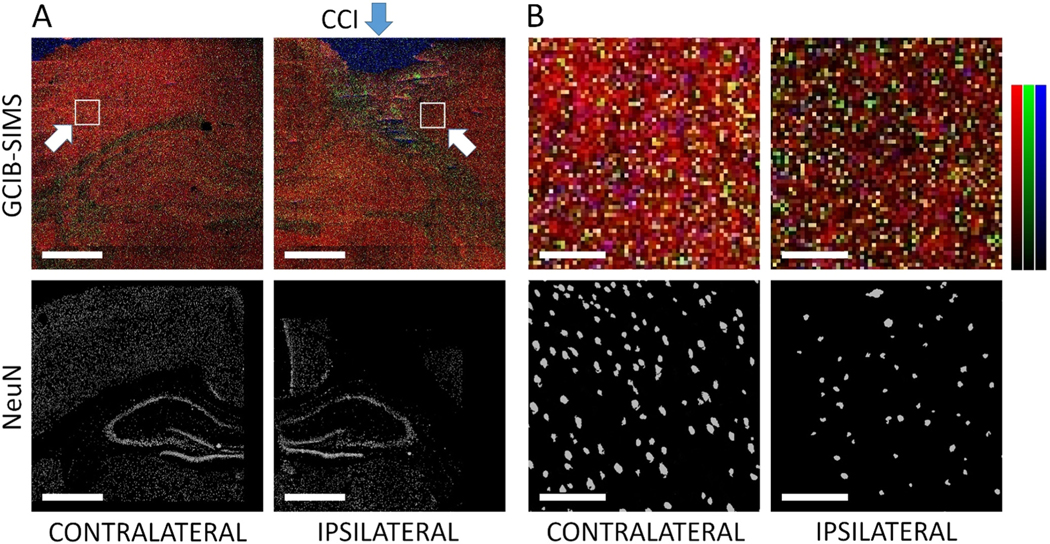
Peroxidized phosphatidylethanolamine (PEox) species have been identified by liquid chromatography mass spectrometry (LC-MS) as predictive biomarkers of ferroptosis, a new program of regulated cell death. However, the presence and subcellular distribution of PEox in specific cell types and tissues have not been directly detected by imaging protocols. By applying gas cluster ion beam secondary ion mass spectrometry (GCIB-SIMS) imaging with a 70 keV (H O) (n>28 000) cluster ion beam, we were able to map PEox with 1.2 μm spatial resolution at the single cell/subcellular level in ferroptotic H9c2 cardiomyocytes and cortical/hippocampal neurons after traumatic brain injury. Application of this protocol affords visualization of physiologically relevant levels of very low abundance (20 pmol μmol lipid) peroxidized lipids in subcellular compartments and their accumulation in disease conditions.
过氧磷脂酰乙醇胺 (PEox) 物种已通过液相色谱-质谱联用 (LC-MS) 被鉴定为铁死亡的预测性生物标志物,铁死亡是一种新的细胞程序性死亡方式。然而,过氧磷脂酰乙醇胺在特定细胞类型和组织中的存在和亚细胞分布尚未通过成像方案直接检测到。通过应用具有 70keV (H O) (n>28000) 团簇离子束的气体团簇离子束二次离子质谱 (GCIB-SIMS) 成像,我们能够以 1.2μm 的空间分辨率在铁死亡的 H9c2 心肌细胞和创伤性脑损伤后的皮质/海马神经元中单细胞/亚细胞水平上对 PEox 进行成像。该方案的应用可实现对亚细胞区室中生理相关低丰度(20pmolμmol 脂质)过氧化脂质的可视化,并可观察到其在疾病状态下的积累。