Department of Chemistry, Pennsylvania State University , State College, Pennsylvania 16802, United States.
Department of Environmental and Occupational Health, University of Pittsburgh , Pittsburgh, Pennsylvania 15260, United States.
Anal Chem. 2017 Apr 18;89(8):4611-4619. doi: 10.1021/acs.analchem.7b00164. Epub 2017 Mar 29.
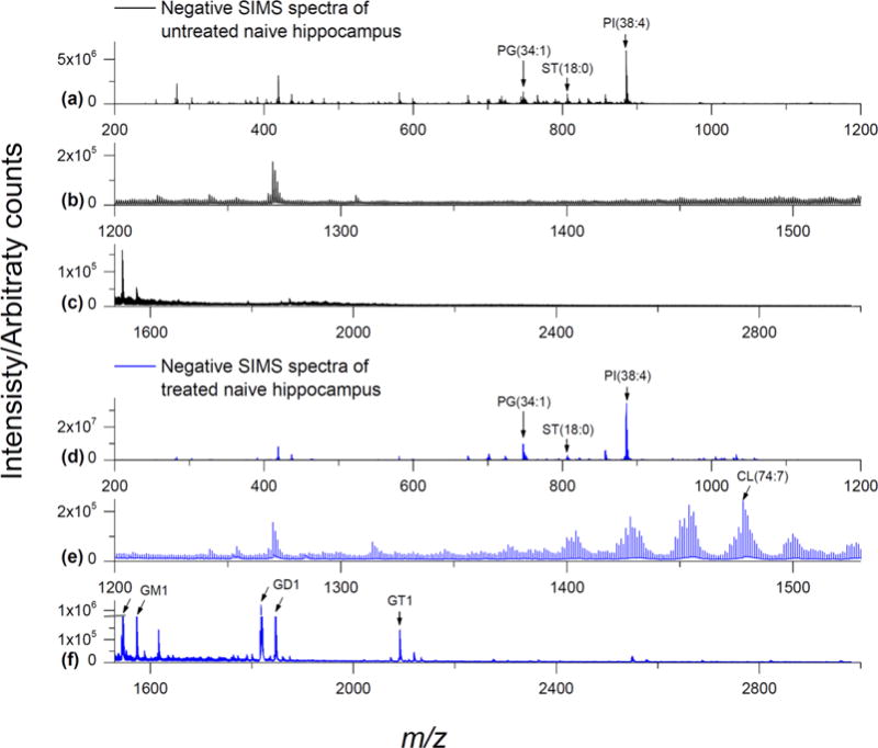
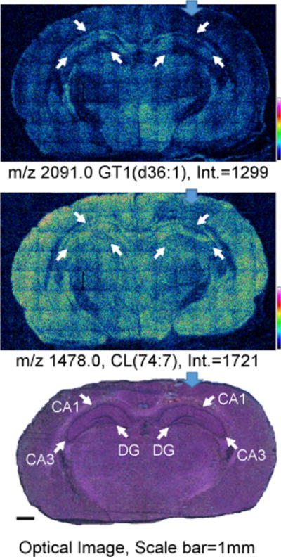
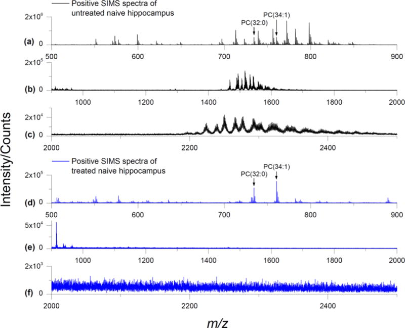
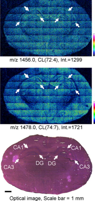
Gas cluster ion beam-secondary ion mass spectrometry (GCIB-SIMS) has shown the full potential of mapping intact lipids in biological systems with better than 10 μm lateral resolution. This study investigated further the capability of GCIB-SIMS in imaging high-mass signals from intact cardiolipin (CL) and gangliosides in normal brain and the effect of a controlled cortical impact model (CCI) of traumatic brain injury (TBI) on their distribution. A combination of enzymatic and chemical treatments was employed to suppress the signals from the most abundant phospholipids (phosphatidylcholine (PC) and phosphatidylethanolamine (PE)) and enhance the signals from the low-abundance CLs and gangliosides to allow their GCIB-SIMS detection at 8 and 16 μm spatial resolution. Brain CLs have not been observed previously using other contemporary imaging mass spectrometry techniques at better than 50 μm spatial resolution. High-resolution images of naive and injured brain tissue facilitated the comparison of CL species across three multicell layers in the CA1, CA3, and DG regions of the hippocampus. GCIB-SIMS also reliably mapped losses of oxidizable polyunsaturated CL species (but not the oxidation-resistant saturated and monounsaturated gangliosides) to regions including the CA1 and CA3 of the hippocampus after CCI. This work extends the detection range for SIMS measurements of intact lipids to above m/z 2000, bridging the mass range gap compared with MALDI. Further advances in high-resolution SIMS of CLs, with the potential for single cell or supra-cellular imaging, will be essential for the understanding of CL's functional and structural organization in normal and injured brain.
气体团簇离子束-二次离子质谱(GCIB-SIMS)已显示出在具有优于 10μm 横向分辨率的生物系统中对完整脂质进行映射的全部潜力。本研究进一步研究了 GCIB-SIMS 在成像完整心磷脂(CL)和神经节苷脂的高质量信号方面的能力,以及受控皮质撞击模型(CCI)对创伤性脑损伤(TBI)对其分布的影响。采用酶和化学处理的组合来抑制最丰富的磷脂(磷脂酰胆碱(PC)和磷脂酰乙醇胺(PE))的信号,并增强低丰度 CL 和神经节苷脂的信号,以允许在 8μm 和 16μm 空间分辨率下对其进行 GCIB-SIMS 检测。使用其他当代成像质谱技术,在优于 50μm 空间分辨率的情况下,此前尚未观察到脑 CL。对未受伤和受伤脑组织的高分辨率图像有助于比较海马 CA1、CA3 和 DG 区三个多细胞层中 CL 物种的分布。GCIB-SIMS 还可靠地绘制了氧化可还原多不饱和 CL 物种(而不是氧化抗性饱和和单不饱和神经节苷脂)在 CCI 后包括海马 CA1 和 CA3 在内的区域的损失。这项工作将 SIMS 对完整脂质的测量范围扩展到 m/z 2000 以上,与 MALDI 相比,弥合了质量范围的差距。CL 高分辨率 SIMS 的进一步发展,具有单细胞或超细胞成像的潜力,对于理解 CL 在正常和受伤大脑中的功能和结构组织至关重要。