• 文献检索
  • 文档翻译
  • 深度研究
  • 学术资讯
  • Suppr Zotero 插件Zotero 插件
  • 邀请有礼
  • 套餐&价格
  • 历史记录
应用&插件
Suppr Zotero 插件Zotero 插件浏览器插件Mac 客户端Windows 客户端微信小程序
定价
高级版会员购买积分包购买API积分包
服务
文献检索文档翻译深度研究API 文档MCP 服务
关于我们
关于 Suppr公司介绍联系我们用户协议隐私条款
关注我们

Suppr 超能文献

核心技术专利:CN118964589B侵权必究
粤ICP备2023148730 号-1Suppr @ 2026

文献检索

告别复杂PubMed语法,用中文像聊天一样搜索,搜遍4000万医学文献。AI智能推荐,让科研检索更轻松。

立即免费搜索

文件翻译

保留排版,准确专业,支持PDF/Word/PPT等文件格式,支持 12+语言互译。

免费翻译文档

深度研究

AI帮你快速写综述,25分钟生成高质量综述,智能提取关键信息,辅助科研写作。

立即免费体验

眼部前段成像技术的进展

Advances in Imaging Technology of Anterior Segment of the Eye.

作者信息

Han Sang Beom, Liu Yu-Chi, Mohamed-Noriega Karim, Mehta Jodhbir S

机构信息

Department of Ophthalmology, Kangwon National University School of Medicine, Kangwon National University Hospital, Chuncheon, Republic of Korea.

Singapore National Eye Centre, Singapore, Singapore.

出版信息

J Ophthalmol. 2021 Feb 23;2021:9539765. doi: 10.1155/2021/9539765. eCollection 2021.

DOI:10.1155/2021/9539765
PMID:33688432
原文链接:https://pmc.ncbi.nlm.nih.gov/articles/PMC7925029/
Abstract

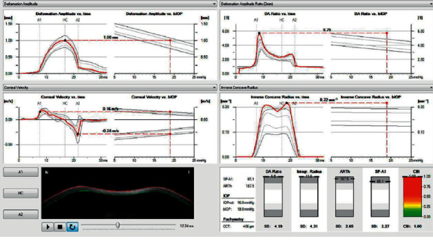
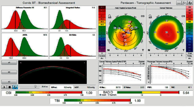
Advances in imaging technology and computer science have allowed the development of newer assessment of the anterior segment, including Corvis ST, Brillouin microscopy, ultrahigh-resolution optical coherence tomography, and artificial intelligence. They enable accurate and precise assessment of structural and biomechanical alterations associated with anterior segment disorders. This review will focus on these 4 new techniques, and a brief overview of these modalities will be introduced. The authors will also discuss the recent advances in research regarding these techniques and potential application of these techniques in clinical practice. Many studies on these modalities have reported promising results, indicating the potential for more detailed comprehensive understanding of the anterior segment tissues.

摘要

成像技术和计算机科学的进步推动了眼前节评估新技术的发展,包括Corvis ST、布里渊显微镜、超高分辨率光学相干断层扫描以及人工智能。它们能够对与眼前节疾病相关的结构和生物力学改变进行准确而精确的评估。本综述将聚焦于这四种新技术,并对这些技术进行简要概述。作者还将讨论这些技术的最新研究进展以及它们在临床实践中的潜在应用。许多关于这些技术的研究都报告了令人鼓舞的结果,表明有可能更详细、全面地了解眼前节组织。

https://cdn.ncbi.nlm.nih.gov/pmc/blobs/b8dd/7925029/5d0963a6e78a/joph2021-9539765.002.jpg
https://cdn.ncbi.nlm.nih.gov/pmc/blobs/b8dd/7925029/dd321b3c7383/joph2021-9539765.001.jpg
https://cdn.ncbi.nlm.nih.gov/pmc/blobs/b8dd/7925029/5d0963a6e78a/joph2021-9539765.002.jpg
https://cdn.ncbi.nlm.nih.gov/pmc/blobs/b8dd/7925029/dd321b3c7383/joph2021-9539765.001.jpg
https://cdn.ncbi.nlm.nih.gov/pmc/blobs/b8dd/7925029/5d0963a6e78a/joph2021-9539765.002.jpg

相似文献

1
Advances in Imaging Technology of Anterior Segment of the Eye.眼部前段成像技术的进展
J Ophthalmol. 2021 Feb 23;2021:9539765. doi: 10.1155/2021/9539765. eCollection 2021.
2
Anterior segment imaging: ultrasound biomicroscopy and anterior segment optical coherence tomography.眼前节成像:超声生物显微镜检查和眼前节光学相干断层扫描
Curr Opin Ophthalmol. 2008 Mar;19(2):115-21. doi: 10.1097/ICU.0b013e3282f40bba.
3
Anterior segment optical coherence tomography: its application in clinical practice and experimental models of disease.眼前节光学相干断层扫描:其在临床实践及疾病实验模型中的应用
Clin Exp Optom. 2019 May;102(3):208-217. doi: 10.1111/cxo.12835. Epub 2018 Oct 1.
4
Ultra-high resolution optical coherence tomography for imaging the anterior segment of the eye.用于眼部前段成像的超高分辨率光学相干断层扫描技术
Ophthalmic Surg Lasers Imaging. 2011 Jul;42 Suppl:S15-27. doi: 10.3928/15428877-20110627-02.
5
Applications of Anterior Segment Optical Coherence Tomography in Cornea and Ocular Surface Diseases.眼前节光学相干断层扫描在角膜和眼表疾病中的应用
J Ophthalmol. 2016;2016:4971572. doi: 10.1155/2016/4971572. Epub 2016 Sep 19.
6
Anterior segment optical coherence tomography.眼前节光学相干断层扫描。
Prog Retin Eye Res. 2018 Sep;66:132-156. doi: 10.1016/j.preteyeres.2018.04.002. Epub 2018 Apr 7.
7
Biomechanical diagnostics of the cornea.角膜的生物力学诊断
Eye Vis (Lond). 2020 Feb 5;7:9. doi: 10.1186/s40662-020-0174-x. eCollection 2020.
8
Optical coherence tomography angiography for the anterior segment.眼前节光学相干断层扫描血管造影术
Eye Vis (Lond). 2019 Feb 1;6:4. doi: 10.1186/s40662-019-0129-2. eCollection 2019.
9
Real-time optical coherence tomography of the anterior segment at 1310 nm.1310纳米波长下眼前节的实时光学相干断层扫描
Arch Ophthalmol. 2001 Aug;119(8):1179-85. doi: 10.1001/archopht.119.8.1179.
10
Anterior segment optical coherence tomography.眼前节光学相干断层扫描
Semin Ophthalmol. 2010 Sep-Nov;25(5-6):317-23. doi: 10.3109/08820538.2010.518473.

引用本文的文献

1
Clinical Applications of Artificial Intelligence in Corneal Diseases.人工智能在角膜疾病中的临床应用
Vision (Basel). 2025 Aug 18;9(3):71. doi: 10.3390/vision9030071.
2
Long-Term Clinical and Structural Outcomes Following Iris-Claw IOL Exchange for Dislocated Intraocular Lenses.虹膜爪型人工晶状体置换脱位人工晶状体后的长期临床和结构转归
J Clin Med. 2025 May 9;14(10):3306. doi: 10.3390/jcm14103306.
3
Integration of Artificial Intelligence into the Approach for Diagnosis and Monitoring of Dry Eye Disease.人工智能在干眼疾病诊断与监测方法中的整合

本文引用的文献

1
Insights into Systemic Disease through Retinal Imaging-Based Oculomics.基于视网膜成像的眼科学洞察全身性疾病。
Transl Vis Sci Technol. 2020 Feb 12;9(2):6. doi: 10.1167/tvst.9.2.6.
2
Deep learning segmentation for optical coherence tomography measurements of the lower tear meniscus.用于下泪液半月皱襞光学相干断层扫描测量的深度学习分割
Biomed Opt Express. 2020 Feb 20;11(3):1539-1554. doi: 10.1364/BOE.386228. eCollection 2020 Mar 1.
3
Quantitative and qualitative evaluation of deep learning automatic segmentations of corneal endothelial cell images of reduced image quality obtained following cornea transplant.
Diagnostics (Basel). 2022 Dec 14;12(12):3167. doi: 10.3390/diagnostics12123167.
4
Assessment of corneal biomechanics in anisometropia using Scheimpflug technology.使用Scheimpflug技术评估屈光参差患者的角膜生物力学。
Front Bioeng Biotechnol. 2022 Oct 4;10:994353. doi: 10.3389/fbioe.2022.994353. eCollection 2022.
5
Application of Intraoperative Optical Coherence Tomography Technology in Anterior Segment Surgery.术中光学相干断层扫描技术在前节手术中的应用
J Ophthalmol. 2022 Apr 8;2022:1568406. doi: 10.1155/2022/1568406. eCollection 2022.
对角膜移植后获得的图像质量降低的角膜内皮细胞图像的深度学习自动分割进行定量和定性评估。
J Med Imaging (Bellingham). 2020 Jan;7(1):014503. doi: 10.1117/1.JMI.7.1.014503. Epub 2020 Feb 14.
4
Biomechanical diagnostics of the cornea.角膜的生物力学诊断
Eye Vis (Lond). 2020 Feb 5;7:9. doi: 10.1186/s40662-020-0174-x. eCollection 2020.
5
Comparison of Corneal Biomechanical Properties among Axial Myopic, Nonaxial Myopic, and Nonmyopic Eyes.轴向近视、非轴向近视与非近视眼中角膜生物力学特性的比较。
Biomed Res Int. 2020 Jan 3;2020:8618615. doi: 10.1155/2020/8618615. eCollection 2020.
6
Automatic Corneal Ulcer Segmentation Combining Gaussian Mixture Modeling and Otsu Method.结合高斯混合模型和大津法的自动角膜溃疡分割
Annu Int Conf IEEE Eng Med Biol Soc. 2019 Jul;2019:6298-6301. doi: 10.1109/EMBC.2019.8857522.
7
Convolutional neural network-based regression for biomarker estimation in corneal endothelium microscopy images.基于卷积神经网络的角膜内皮显微镜图像生物标志物估计回归方法
Annu Int Conf IEEE Eng Med Biol Soc. 2019 Jul;2019:876-881. doi: 10.1109/EMBC.2019.8857201.
8
[Automated Cell Counting Using "Deep Learning" in Donor Corneas from Organ Culture Achieves High Precision and Accuracy].[在器官培养的供体角膜中使用“深度学习”进行自动细胞计数可实现高精度和准确性]
Klin Monbl Augenheilkd. 2019 Dec;236(12):1407-1412. doi: 10.1055/a-1023-4339. Epub 2019 Dec 5.
9
corneal endothelium imaging using ultrahigh resolution OCT.使用超高分辨率光学相干断层扫描技术进行角膜内皮成像
Biomed Opt Express. 2019 Oct 11;10(11):5675-5686. doi: 10.1364/BOE.10.005675. eCollection 2019 Nov 1.
10
A new method for detecting the outer corneal contour in images from an ultra-fast Scheimpflug camera.一种用于检测超快速 Scheimpflug 相机图像中外眼角膜轮廓的新方法。
Biomed Eng Online. 2019 Dec 3;18(1):115. doi: 10.1186/s12938-019-0735-1.