Kiuchi Kunihiko, Fukuzawa Koji, Nogami Munenobu, Watanabe Yoshiaki, Takami Mitsuru, Mori Shumpei, Shimoyama Shinsuke, Negi Noriyuki, Kyotani Katsusuke, Hirata Ken-Ichi
Section of Arrhythmia, Division of Cardiovascular Medicine, Department of Internal Medicine, Kobe University Graduate School of Medicine Kobe Japan.
Department of Radiology, Kobe University Graduate School of Medicine Kobe Japan.
Circ Rep. 2020 Feb 13;1(3):149-152. doi: 10.1253/circrep.CR-19-0003.
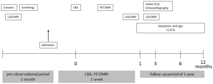
Atrial fibrosis and inflammation play important roles in perpetuating and initiating atrial fibrillation (AF). Although the fibrotic area can be visualized as a delayed enhancement area on late gadolinium enhancement magnetic resonance imaging (LGE-MRI), atrial inflammation has not yet been visualized on any imaging modality. We describe the protocol for a feasibility study to visualize atrial inflammation on positron emission tomography/MRI (PET/MRI). This is a single-arm, prospective, open-label proof-of concept trial, involving AF patients after cryoballoon ablation (CBA). A total of 30 paroxysmal AF patients will be enrolled and undergo simultaneous PET/MRI for the assessment of regional F-fluorodeoxyglucose (F-FDG) uptake 1 day after the CBA. Furthermore, LGE-MRI will be performed before CBA, and at 1 and 4 weeks after assessing the regional LGE area. The main outcome measures will be (1) the feasibility of imaging inflammation in the left atrium on PET/MRI; and (2) the safety of the intervention. There are few data on the visualization of atrial inflammation using PET/MRI. Establishing the visualization methodology will contribute to elucidating the fundamental histopathologic findings of the progress to fibrosis, and to the planning and execution of a larger definitive trial to test the usefulness of PET/MRI.
心房纤维化和炎症在心房颤动(AF)的持续和起始过程中发挥着重要作用。虽然纤维化区域在钆延迟增强磁共振成像(LGE-MRI)上可表现为延迟强化区域,但目前尚无任何成像方式能够显示心房炎症。我们描述了一项关于在正电子发射断层扫描/磁共振成像(PET/MRI)上显示心房炎症的可行性研究方案。这是一项单臂、前瞻性、开放标签的概念验证试验,纳入接受冷冻球囊消融(CBA)后的AF患者。总共将招募30例阵发性AF患者,并在CBA后1天接受同步PET/MRI检查,以评估局部F-氟脱氧葡萄糖(F-FDG)摄取情况。此外,将在CBA前以及评估局部LGE区域后的1周和4周进行LGE-MRI检查。主要观察指标将包括:(1)PET/MRI显示左心房炎症的可行性;(2)干预措施的安全性。关于使用PET/MRI显示心房炎症的数据很少。建立可视化方法将有助于阐明向纤维化进展的基本组织病理学发现,并有助于规划和开展一项更大规模的确定性试验,以检验PET/MRI的实用性。