Department of Cardiology, The First People's Hospital of Shangqiu, China.
Department of Cardiology, The First Affiliated Hospital of Zhengzhou University, China.
J Immunol Res. 2022 May 12;2022:3647817. doi: 10.1155/2022/3647817. eCollection 2022.
To analyze the role of PD-1/PD-L1 signaling pathway in regulating T cell activation and secretion of proinflammatory factors in atrial fibrillation.
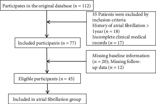
Forty-five patients with atrial fibrillation admitted to the cardiology department of our hospital from July 2019 to March 2021 were selected to be included in the atrial fibrillation group, and another 45 healthy volunteers were selected as the control group to compare the changes of T cell CD69 and human leukocyte antigen-DR (HLA-DR) expression in the peripheral blood of the two study groups; compare the changes of programmed death factor-1 on CD4+ and CD8+ lymphocytes in the peripheral blood of the two groups (PD-1) expression changes and PD-L1 and PD-L2 expression changes on peripheral blood myeloid dendritic cells (mDCs) cells; compare the changes of interleukin-2, interleukin-6, interleukin-10, and interleukin-17A (IL-2, IL-6, IL-10, and IL-17), tumor necrosis factor (TNF), and interferon gamma (IFN-) concentrations on peripheral blood inflammatory factors in the two groups; and isolate the two groups of peripheral blood mDCs cells; interferon upregulated PD-L1 expression in the cells and analyzed the effect of PD-L1 expression on the ability of mDCs to stimulate T cells to secrete cytokines.
The positive expression rates of CD69 and HLA-DR on peripheral blood CD3+ T lymphocytes were significantly higher in patients in the atrial fibrillation group than in the control group, and the differences were statistically significant ( < 0.01). The positive expression rate of PD-1 on CD4+ lymphocytes was significantly lower in patients in the atrial fibrillation group than in the control group ( < 0.01). There was no statistically significant difference between the two groups in terms of PD-1 positive expression rate on CD8+ lymphocytes ( > 0.05). The positive expression rate of PD-L1 on mDCs cells was significantly lower in patients in the atrial fibrillation group than in the control group ( < 0.01), and there was no statistically significant difference between the two groups in the positive expression rate of PD-L2 on mDCs cells, PD-L1, and PD-L2 on CD4+ and CD8+ T cells ( > 0.05). The concentrations of IL-2, IL-6, IL-10, and IFN- in peripheral blood were significantly higher in patients in the atrial fibrillation group than in the control group ( < 0.05), and there was no statistically significant difference in the comparison of IL-17A and TNF concentrations in peripheral blood between the two groups ( > 0.05). In the atrial fibrillation group, the ability of mDCs to stimulate T cells to secrete IL-2 and IFN- was significantly higher, and the ability to secrete IL-10 was significantly lower compared with the control group (P < 0.05). After interferon upregulated PD-L1 expression in cells, the ability of mDCs to stimulate T cells to secrete IL-2, IL-10, and IFN- cytokines was reversed in patients in the atrial fibrillation group, and the differences compared with the control group were not statistically significant ( > 0.05).
PD-1/PD-L1 signaling pathway may play an immunomodulatory role in the pathogenesis of atrial fibrillation by promoting increased secretion of inflammatory factors through regulating T cell activation.
分析 PD-1/PD-L1 信号通路在调节心房颤动中 T 细胞激活和促炎因子分泌中的作用。
选取 2019 年 7 月至 2021 年 3 月我院心内科收治的 45 例心房颤动患者纳入心房颤动组,另选取同期 45 例健康志愿者纳入对照组,比较两组外周血 T 细胞 CD69 和人类白细胞抗原-DR(HLA-DR)表达的变化;比较两组外周血 CD4+和 CD8+淋巴细胞程序性死亡因子-1(PD-1)表达变化及外周血髓样树突状细胞(mDCs)程序性死亡配体-1(PD-L1)和 PD-L2 表达变化;比较两组外周血炎症因子白细胞介素-2(IL-2)、白细胞介素-6(IL-6)、白细胞介素-10(IL-10)、白细胞介素-17A(IL-17A)、肿瘤坏死因子(TNF)和干扰素-γ(IFN-γ)浓度的变化;分离两组外周血 mDCs 细胞;用干扰素上调细胞中 PD-L1 表达,并分析 PD-L1 表达对 mDCs 刺激 T 细胞分泌细胞因子能力的影响。
心房颤动组患者外周血 CD3+T 淋巴细胞 CD69 和 HLA-DR 的阳性表达率明显高于对照组,差异有统计学意义( < 0.01)。心房颤动组患者 CD4+淋巴细胞 PD-1 阳性表达率明显低于对照组( < 0.01)。两组 CD8+淋巴细胞 PD-1 阳性表达率比较,差异无统计学意义( > 0.05)。心房颤动组患者 mDCs 细胞 PD-L1 阳性表达率明显低于对照组( < 0.01),两组 mDCs 细胞 PD-L2 阳性表达率、CD4+和 CD8+T 细胞 PD-L1 和 PD-L2 阳性表达率比较,差异无统计学意义( > 0.05)。心房颤动组患者外周血 IL-2、IL-6、IL-10 和 IFN-γ 浓度明显高于对照组( < 0.05),两组外周血 IL-17A 和 TNF 浓度比较,差异无统计学意义( > 0.05)。与对照组比较,心房颤动组 mDCs 刺激 T 细胞分泌 IL-2 和 IFN-γ 的能力明显升高,分泌 IL-10 的能力明显降低(P < 0.05)。用干扰素上调细胞中 PD-L1 表达后,心房颤动组 mDCs 刺激 T 细胞分泌 IL-2、IL-10 和 IFN-γ 细胞因子的能力被逆转,与对照组比较差异无统计学意义( > 0.05)。
PD-1/PD-L1 信号通路可能通过调节 T 细胞激活促进促炎因子的大量分泌,在心房颤动的发病机制中发挥免疫调节作用。