• 文献检索
  • 文档翻译
  • 深度研究
  • 学术资讯
  • Suppr Zotero 插件Zotero 插件
  • 邀请有礼
  • 套餐&价格
  • 历史记录
应用&插件
Suppr Zotero 插件Zotero 插件浏览器插件Mac 客户端Windows 客户端微信小程序
定价
高级版会员购买积分包购买API积分包
服务
文献检索文档翻译深度研究API 文档MCP 服务
关于我们
关于 Suppr公司介绍联系我们用户协议隐私条款
关注我们

Suppr 超能文献

核心技术专利:CN118964589B侵权必究
粤ICP备2023148730 号-1Suppr @ 2026

文献检索

告别复杂PubMed语法,用中文像聊天一样搜索,搜遍4000万医学文献。AI智能推荐,让科研检索更轻松。

立即免费搜索

文件翻译

保留排版,准确专业,支持PDF/Word/PPT等文件格式,支持 12+语言互译。

免费翻译文档

深度研究

AI帮你快速写综述,25分钟生成高质量综述,智能提取关键信息,辅助科研写作。

立即免费体验

多壳扩散 MRI 显示多发性硬化症早期微观结构白质异常的证据。

Evidence of early microstructural white matter abnormalities in multiple sclerosis from multi-shell diffusion MRI.

机构信息

Instituto de Neurociencias de Alicante (CSIC-UMH), San Juan de Alicante, Spain; Cardiff University Brain Research Imaging Centre (CUBRIC), Cardiff University, Cardiff, UK.

Department of Clinical Neuroscience, Karolinska Institutet, Stockholm, Sweden; Department of Radiology, Karolinska University Hospital, Stockholm, Sweden; Athinoula A. Martinos Center for Biomedical Imaging and Harvard Medical School, Boston, MA, USA.

出版信息

Neuroimage Clin. 2019;22:101699. doi: 10.1016/j.nicl.2019.101699. Epub 2019 Jan 30.

DOI:10.1016/j.nicl.2019.101699
PMID:30739842
原文链接:https://pmc.ncbi.nlm.nih.gov/articles/PMC6370560/
Abstract

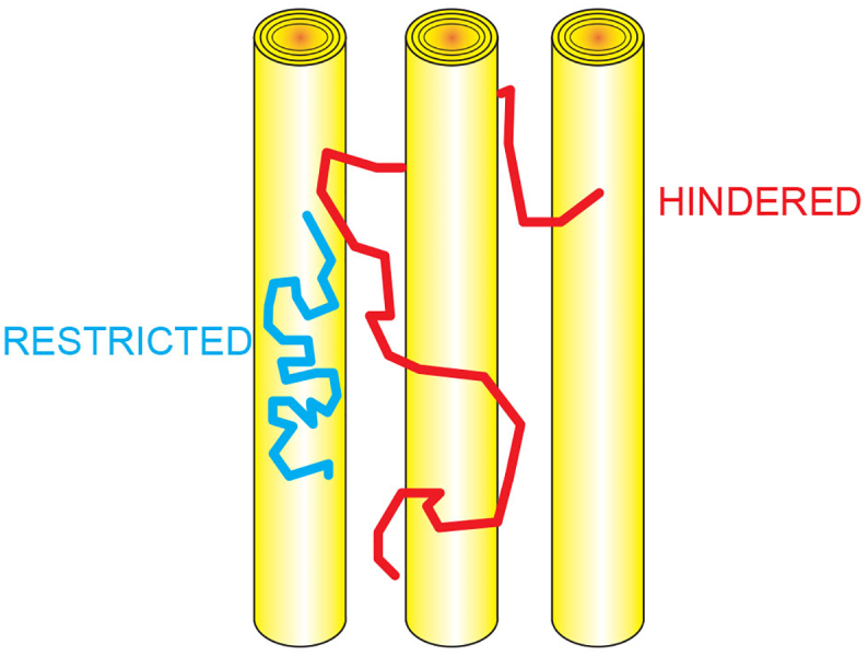
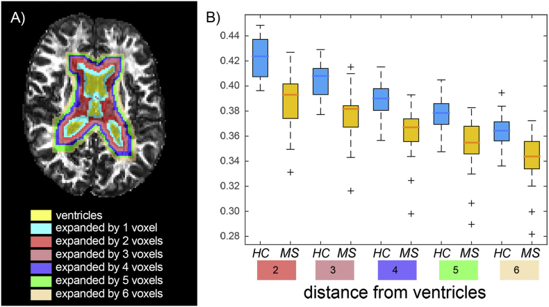
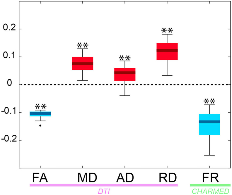
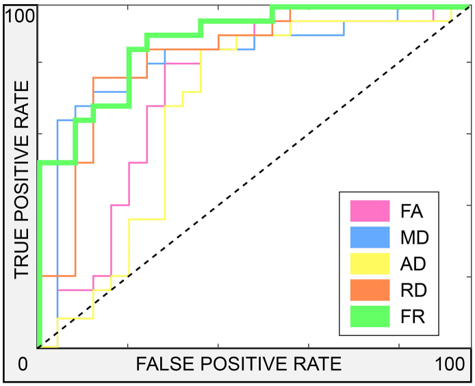
Irreversible white matter (WM) damage, including severe demyelination and axonal loss, is a main determinant of long-term disability in multiple sclerosis (MS). Non-invasive detection of changes in microstructural WM integrity in the disease is challenging since commonly used imaging metrics lack the necessary sensitivity, especially in the early phase of the disease. This study aims at assessing microstructural WM abnormalities in early-stage MS by using ultra-high gradient strength multi-shell diffusion MRI and the restricted signal fraction (FR) from the Composite Hindered and Restricted Model of Diffusion (CHARMED), a metric sensitive to the volume fraction of axons. In 22 early MS subjects (disease duration ≤5 years) and 15 age-matched healthy controls, restricted fraction estimates were obtained through the CHARMED model along with conventional Diffusion Tensor Imaging (DTI) metrics. All imaging parameters were compared cross-sectionally between the MS subjects and controls both in WM lesions and normal-appearing white matter (NAWM). We found a significant reduction in FR focally in WM lesions and widespread in the NAWM in MS patients relative to controls (corrected p < .05). Signal fraction changes in NAWM were not driven by perilesional tissue, nor were they influenced by proximity to the ventricles, challenging the hypothesis of an outside-in pathological process driven by CSF-mediated immune cytotoxic factors. No significant differences were found in conventional DTI parameters. In a cross-validated classification task, FR showed the largest effect size and outperformed all other diffusion imaging metrics in discerning lesions from contralateral NAWM. Taken together, our data provide evidence for the presence of widespread microstructural changes in the NAWM in early MS stages that are, at least in part, unrelated to focal demyelinating lesions. Interestingly, these pathological changes were not yet detectable by conventional diffusion imaging at this early disease stage, highlighting the sensitivity and value of multi-shell diffusion imaging for better characterizing axonal microstructure in MS.

摘要

不可逆的脑白质(WM)损伤,包括严重的脱髓鞘和轴突丢失,是多发性硬化症(MS)长期残疾的主要决定因素。由于常用的成像指标缺乏必要的敏感性,特别是在疾病的早期阶段,因此很难无创地检测疾病中微观结构 WM 完整性的变化。本研究旨在通过使用超高梯度强度多壳扩散 MRI 和受限信号分数(FR)来评估早期 MS 中的微观结构 WM 异常,受限信号分数是一种对轴突体积分数敏感的扩散复合受限和受限模型(CHARMED)的指标。在 22 名早期 MS 患者(病程≤5 年)和 15 名年龄匹配的健康对照者中,通过 CHARMED 模型获得受限分数估计值,以及常规扩散张量成像(DTI)指标。在 MS 患者和对照组的 WM 病变和正常表现的白质(NAWM)之间,比较了所有成像参数的横截面差异。与对照组相比,MS 患者的 WM 病变和 NAWM 中存在明显的 FR 局灶性降低和广泛降低(校正后 p<0.05)。NAWM 中的信号分数变化不是由病变周围组织驱动的,也不受与脑室接近程度的影响,这对 CSF 介导的免疫细胞毒性因子驱动的由外向内的病理过程假说提出了挑战。在常规 DTI 参数中未发现显著差异。在交叉验证分类任务中,FR 显示出最大的效应量,并且在辨别病变与对侧 NAWM 方面优于所有其他扩散成像指标。综上所述,我们的数据提供了证据,证明在早期 MS 阶段,NAWM 中存在广泛的微观结构变化,至少部分与局灶性脱髓鞘病变无关。有趣的是,在疾病的早期阶段,这些病理变化还不能通过常规扩散成像检测到,这凸显了多壳扩散成像的敏感性和价值,有助于更好地描述 MS 中的轴突微观结构。

https://cdn.ncbi.nlm.nih.gov/pmc/blobs/b864/6370560/c3eaa8ebb079/gr7.jpg
https://cdn.ncbi.nlm.nih.gov/pmc/blobs/b864/6370560/2704c2d50c75/gr1.jpg
https://cdn.ncbi.nlm.nih.gov/pmc/blobs/b864/6370560/c5dc319b848a/gr2.jpg
https://cdn.ncbi.nlm.nih.gov/pmc/blobs/b864/6370560/fd52bdfed110/gr3.jpg
https://cdn.ncbi.nlm.nih.gov/pmc/blobs/b864/6370560/fe49c49b72a5/gr4.jpg
https://cdn.ncbi.nlm.nih.gov/pmc/blobs/b864/6370560/71989fad1b56/gr5.jpg
https://cdn.ncbi.nlm.nih.gov/pmc/blobs/b864/6370560/8f0bdd0610ae/gr6.jpg
https://cdn.ncbi.nlm.nih.gov/pmc/blobs/b864/6370560/c3eaa8ebb079/gr7.jpg

相似文献

1
Evidence of early microstructural white matter abnormalities in multiple sclerosis from multi-shell diffusion MRI.多壳扩散 MRI 显示多发性硬化症早期微观结构白质异常的证据。
Neuroimage Clin. 2019;22:101699. doi: 10.1016/j.nicl.2019.101699. Epub 2019 Jan 30.
2
Evidence for Progressive Microstructural Damage in Early Multiple Sclerosis by Multi-Shell Diffusion Magnetic Resonance Imaging.多壳层弥散磁共振成像显示早期多发性硬化症的进行性微观结构损伤证据。
Neuroscience. 2019 Apr 1;403:27-34. doi: 10.1016/j.neuroscience.2019.01.022. Epub 2019 Jan 29.
3
Axonal damage in the optic radiation assessed by white matter tract integrity metrics is associated with retinal thinning in multiple sclerosis.轴突损伤在评估视辐射中的白质束完整性指标与多发性硬化症中的视网膜变薄有关。
Neuroimage Clin. 2020;27:102293. doi: 10.1016/j.nicl.2020.102293. Epub 2020 May 26.
4
In vivo characterization of cortical and white matter neuroaxonal pathology in early multiple sclerosis.早期多发性硬化症中皮质和白质神经轴突病变的体内特征分析
Brain. 2017 Nov 1;140(11):2912-2926. doi: 10.1093/brain/awx247.
5
A systematic review of microstructural abnormalities in multiple sclerosis detected with NODDI and DTI models of diffusion-weighted magnetic resonance imaging.基于磁共振弥散加权成像 NODDI 和 DTI 模型检测多发性硬化的微观结构异常的系统评价。
Magn Reson Imaging. 2023 Dec;104:61-71. doi: 10.1016/j.mri.2023.09.010. Epub 2023 Sep 27.
6
Imaging G-Ratio in Multiple Sclerosis Using High-Gradient Diffusion MRI and Macromolecular Tissue Volume.使用高梯度弥散 MRI 和大分子组织容积成像技术评估多发性硬化症的 G 比值
AJNR Am J Neuroradiol. 2019 Nov;40(11):1871-1877. doi: 10.3174/ajnr.A6283. Epub 2019 Nov 6.
7
Comparative overview of multi-shell diffusion MRI models to characterize the microstructure of multiple sclerosis lesions and periplaques.多壳扩散 MRI 模型对多发性硬化病变及斑块微结构特征的比较综述。
Neuroimage Clin. 2024;42:103593. doi: 10.1016/j.nicl.2024.103593. Epub 2024 Mar 18.
8
Comparison of diffusion tensor imaging (DTI) tissue characterization parameters in white matter tracts of patients with multiple sclerosis (MS) and neuromyelitis optica spectrum disorder (NMOSD).比较多发性硬化症(MS)和视神经脊髓炎谱系障碍(NMOSD)患者的弥散张量成像(DTI)白质束组织特征参数。
Eur Radiol. 2024 Aug;34(8):5263-5275. doi: 10.1007/s00330-023-10550-1. Epub 2024 Jan 4.
9
NODDI, diffusion tensor microstructural abnormalities and atrophy of brain white matter and gray matter contribute to cognitive impairment in multiple sclerosis.神经突方向离散与密度成像(NODDI)、脑白质和灰质的扩散张量微观结构异常及萎缩导致多发性硬化症的认知障碍。
J Neurol. 2023 Feb;270(2):810-823. doi: 10.1007/s00415-022-11415-1. Epub 2022 Oct 6.
10
Myelin and axon pathology in multiple sclerosis assessed by myelin water and multi-shell diffusion imaging.多发性硬化症的髓鞘和轴突病理通过髓鞘水和多壳扩散成像评估。
Brain. 2021 Jul 28;144(6):1684-1696. doi: 10.1093/brain/awab088.

引用本文的文献

1
A focus on the normal-appearing white and gray matter within the multiple sclerosis brain: a link to smoldering progression.聚焦多发性硬化症患者大脑中外观正常的白质和灰质:与隐匿性进展的关联
Acta Neuropathol. 2025 Aug 10;150(1):16. doi: 10.1007/s00401-025-02923-1.
2
Moderating effects of limbic white matter on verbal memory in older adults with multiple sclerosis.边缘白质对患有多发性硬化症的老年人言语记忆的调节作用。
Neuropsychology. 2025 Jul 24. doi: 10.1037/neu0001023.
3
Differential Expression of PACAP/VIP Receptors in the Post-Mortem CNS White Matter of Multiple Sclerosis Donors.

本文引用的文献

1
Characterizing Microstructural Tissue Properties in Multiple Sclerosis with Diffusion MRI at 7 T and 3 T: The Impact of the Experimental Design.用 7T 和 3T 的弥散 MRI 对多发性硬化症进行微观结构组织特性分析:实验设计的影响。
Neuroscience. 2019 Apr 1;403:17-26. doi: 10.1016/j.neuroscience.2018.03.048. Epub 2018 Apr 7.
2
In vivo characterization of cortical and white matter neuroaxonal pathology in early multiple sclerosis.早期多发性硬化症中皮质和白质神经轴突病变的体内特征分析
Brain. 2017 Nov 1;140(11):2912-2926. doi: 10.1093/brain/awx247.
3
TE dependent Diffusion Imaging (TEdDI) distinguishes between compartmental T relaxation times.
多发性硬化症供体死后中枢神经系统白质中 PACAP/VIP 受体的差异表达。
Int J Mol Sci. 2024 Aug 14;25(16):8850. doi: 10.3390/ijms25168850.
4
Beyond the division of multiple sclerosis into different subgroups: The Concept of Connectomopathy.超越将多发性硬化症分为不同亚组:连接组病的概念。
Caspian J Intern Med. 2024 Summer;15(3):370-373. doi: 10.22088/cjim.15.3.370.
5
Deep learning with diffusion MRI as in vivo microscope reveals sex-related differences in human white matter microstructure.基于扩散 MRI 的深度学习作为活体显微镜揭示了人类白质微观结构中的性别差异。
Sci Rep. 2024 May 14;14(1):9835. doi: 10.1038/s41598-024-60340-y.
6
A translational MRI approach to validate acute axonal damage detection as an early event in multiple sclerosis.一种转化 MRI 方法,用于验证急性轴索损伤检测作为多发性硬化症的早期事件。
Elife. 2024 Jan 9;13:e79169. doi: 10.7554/eLife.79169.
7
Multishell diffusion MRI reveals whole-brain white matter changes in HIV.多壳弥散 MRI 揭示 HIV 患者全脑白质变化。
Hum Brain Mapp. 2023 Oct 15;44(15):5113-5124. doi: 10.1002/hbm.26448. Epub 2023 Aug 30.
8
Proteomics and relationship with axonal pathology in multiple sclerosis: 5-year diffusion tensor imaging study.蛋白质组学与多发性硬化症轴突病理学的关系:5年扩散张量成像研究
Brain Commun. 2023 Jun 13;5(3):fcad183. doi: 10.1093/braincomms/fcad183. eCollection 2023.
9
Neurofilament light chains in serum as biomarkers of axonal damage in early MS lesions: a histological-serological correlative study.血清神经丝轻链作为早期多发性硬化症病变中轴突损伤的生物标志物:一项组织病理学-血清学相关性研究。
J Neurol. 2023 Mar;270(3):1416-1429. doi: 10.1007/s00415-022-11468-2. Epub 2022 Nov 13.
10
An Update on the Measurement of Motor Cerebellar Dysfunction in Multiple Sclerosis.多发性硬化症中运动小脑功能障碍的测量方法更新。
Cerebellum. 2023 Aug;22(4):761-775. doi: 10.1007/s12311-022-01435-y. Epub 2022 Jun 27.
TE 依赖性扩散成像(TEdDI)可区分隔室 T 弛豫时间。
Neuroimage. 2018 Nov 15;182:360-369. doi: 10.1016/j.neuroimage.2017.09.030. Epub 2017 Sep 19.
4
Sensitivity of multi-shell NODDI to multiple sclerosis white matter changes: a pilot study.多壳层神经突方向离散与密度成像(NODDI)对多发性硬化症白质变化的敏感性:一项初步研究。
Funct Neurol. 2017 Apr/Jun;32(2):97-101. doi: 10.11138/fneur/2017.32.2.097.
5
Evaluating fibre orientation dispersion in white matter: Comparison of diffusion MRI, histology and polarized light imaging.评估白质中的纤维方向分散:弥散磁共振成像、组织学和偏振光成像的比较。
Neuroimage. 2017 Aug 15;157:561-574. doi: 10.1016/j.neuroimage.2017.06.001. Epub 2017 Jun 8.
6
Application and evaluation of NODDI in the cervical spinal cord of multiple sclerosis patients.NODDI在多发性硬化症患者颈脊髓中的应用与评估
Neuroimage Clin. 2017 May 17;15:333-342. doi: 10.1016/j.nicl.2017.05.010. eCollection 2017.
7
Relationship of acute axonal damage, Wallerian degeneration, and clinical disability in multiple sclerosis.多发性硬化症中急性轴索损伤、华勒氏变性与临床残疾的关系。
J Neuroinflammation. 2017 Mar 17;14(1):57. doi: 10.1186/s12974-017-0831-8.
8
An abnormal periventricular magnetization transfer ratio gradient occurs early in multiple sclerosis.室周磁化传递率梯度异常在多发性硬化症早期就会出现。
Brain. 2017 Feb;140(2):387-398. doi: 10.1093/brain/aww296. Epub 2017 Jan 2.
9
Neurite density imaging versus imaging of microscopic anisotropy in diffusion MRI: A model comparison using spherical tensor encoding.扩散磁共振成像中神经突密度成像与微观各向异性成像的比较:使用球张量编码的模型对比
Neuroimage. 2017 Feb 15;147:517-531. doi: 10.1016/j.neuroimage.2016.11.053. Epub 2016 Nov 27.
10
"A new imaging modality to non-invasively assess multiple sclerosis pathology".一种用于非侵入性评估多发性硬化症病理学的新型成像方式
J Neuroimmunol. 2017 Mar 15;304:81-85. doi: 10.1016/j.jneuroim.2016.10.002. Epub 2016 Oct 8.