Region Västra Götaland, Department of Clinical Physiology, Sahlgrenska University Hospital, Gothenburg, Sweden.
Department of Electrical Engineering, Chalmers University of Technology, Gothenburg, Sweden.
Eur Radiol Exp. 2021 Mar 11;5(1):11. doi: 10.1186/s41747-021-00210-8.
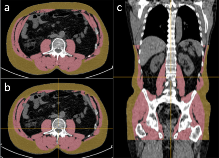
Body composition is associated with survival outcome in oncological patients, but it is not routinely calculated. Manual segmentation of subcutaneous adipose tissue (SAT) and muscle is time-consuming and therefore limited to a single CT slice. Our goal was to develop an artificial-intelligence (AI)-based method for automated quantification of three-dimensional SAT and muscle volumes from CT images.
Ethical approvals from Gothenburg and Lund Universities were obtained. Convolutional neural networks were trained to segment SAT and muscle using manual segmentations on CT images from a training group of 50 patients. The method was applied to a separate test group of 74 cancer patients, who had two CT studies each with a median interval between the studies of 3 days. Manual segmentations in a single CT slice were used for comparison. The accuracy was measured as overlap between the automated and manual segmentations.
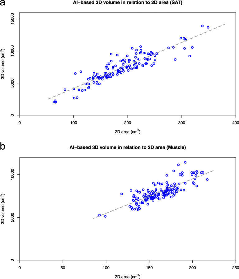
The accuracy of the AI method was 0.96 for SAT and 0.94 for muscle. The average differences in volumes were significantly lower than the corresponding differences in areas in a single CT slice: 1.8% versus 5.0% (p < 0.001) for SAT and 1.9% versus 3.9% (p < 0.001) for muscle. The 95% confidence intervals for predicted volumes in an individual subject from the corresponding single CT slice areas were in the order of ± 20%.
The AI-based tool for quantification of SAT and muscle volumes showed high accuracy and reproducibility and provided a body composition analysis that is more relevant than manual analysis of a single CT slice.
体成分与肿瘤患者的生存结果相关,但目前并未常规计算体成分。手动分割皮下脂肪组织(SAT)和肌肉组织既耗时又费力,因此只能在单个 CT 切片上进行。我们的目标是开发一种基于人工智能(AI)的方法,从 CT 图像自动量化三维 SAT 和肌肉体积。
哥德堡大学和隆德大学伦理委员会批准了本研究。使用 50 例患者 CT 图像的手动分割数据对卷积神经网络进行训练,以分割 SAT 和肌肉组织。该方法应用于 74 例癌症患者的独立测试组,每位患者均接受两次 CT 检查,两次检查的中位间隔为 3 天。使用单个 CT 切片的手动分割进行比较。准确性通过自动化和手动分割之间的重叠来衡量。
AI 方法对 SAT 和肌肉的准确性分别为 0.96 和 0.94。体积的平均差异明显低于单个 CT 切片面积的相应差异:SAT 为 1.8%对 5.0%(p < 0.001),肌肉为 1.9%对 3.9%(p < 0.001)。从相应的单个 CT 切片面积预测个体患者的体积的 95%置信区间在±20%左右。
用于量化 SAT 和肌肉体积的基于 AI 的工具具有很高的准确性和可重复性,并且提供了比手动分析单个 CT 切片更相关的体成分分析。