Morini Raffaella, Bizzotto Matteo, Perrucci Fabio, Filipello Fabia, Matteoli Michela
Laboratory of Pharmacology and Brain Pathology, Neurocenter, Humanitas Clinical and Research Center - IRCCS, Rozzano, Italy.
Department of Biomedical Sciences, Humanitas University, Pieve Emanuele, Italy.
Front Immunol. 2021 Feb 23;12:640937. doi: 10.3389/fimmu.2021.640937. eCollection 2021.
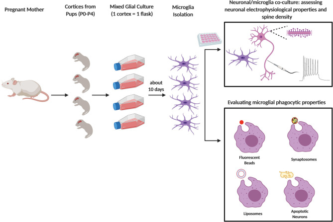
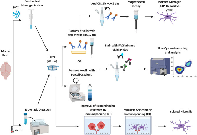
The role of microglia in controlling synapse homeostasis is becoming increasingly recognized by the scientific community. In particular, the microglia-mediated elimination of supernumerary synapses during development lays the basis for the correct formation of neuronal circuits in adulthood, while the possible reactivation of this process in pathological conditions, such as schizophrenia or Alzheimer's Disease, provides a promising target for future therapeutic strategies. The methodological approaches to investigate microglial synaptic engulfment include different and settings. Basic assays, employing isolated microglia and microbeads, apoptotic membranes, liposomes or synaptosomes allow the quantification of the microglia phagocytic abilities, while co-cultures of microglia and neurons, deriving from either WT or genetically modified mice models, provide a relatively manageable setting to investigate the involvement of specific molecular pathways. Further detailed analysis in mice brain is then mandatory to validate the assays as representative for the situation. The present review aims to dissect the main technical approaches to investigate microglia-mediated phagocytosis of neuronal and synaptic substrates in critical developmental time windows.
小胶质细胞在控制突触稳态中的作用日益受到科学界的认可。特别是,小胶质细胞在发育过程中介导多余突触的消除为成年期神经元回路的正确形成奠定了基础,而在诸如精神分裂症或阿尔茨海默病等病理条件下这一过程可能的重新激活为未来的治疗策略提供了一个有前景的靶点。研究小胶质细胞突触吞噬作用的方法学途径包括不同的实验和设置。基本实验采用分离的小胶质细胞和微珠、凋亡膜、脂质体或突触体,可对小胶质细胞的吞噬能力进行定量,而来自野生型或基因改造小鼠模型的小胶质细胞与神经元的共培养提供了一个相对易于管理的环境来研究特定分子途径的参与情况。然后必须在小鼠大脑中进行进一步详细分析,以验证这些实验作为代表实际情况的有效性。本综述旨在剖析在关键发育时间窗口研究小胶质细胞介导的神经元和突触底物吞噬作用的主要技术方法。