Biomedical Research Institute UHasselt Hasselt University Hasselt Belgium.
Laboratory of Cell Biology & Histology Antwerp Centre for Advanced Microscopy (ACAM) University of Antwerp Antwerp Belgium.
J Extracell Vesicles. 2020 Nov;10(1):e12022. doi: 10.1002/jev2.12022. Epub 2020 Nov 25.
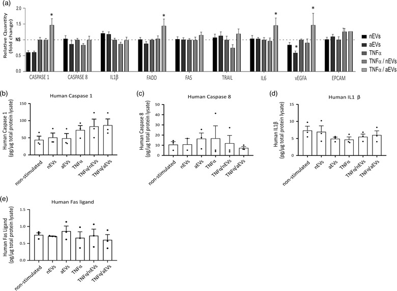
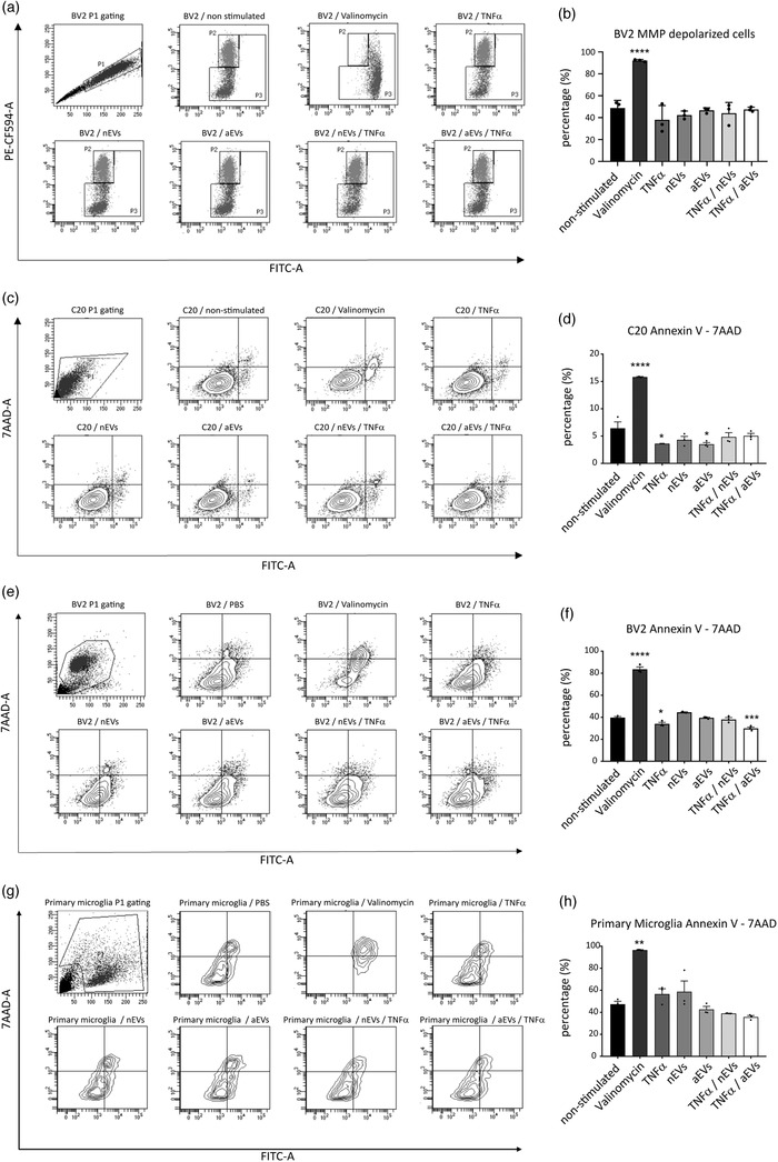
Microglia, the immunocompetent cells of the central nervous system (CNS), play an important role in maintaining cellular homeostasis in the CNS. These cells secrete immunomodulatory factors including nanovesicles and participate in the removal of cellular debris by phagocytosis or autophagy. Accumulating evidence indicates that specifically the cellular exchange of small extracellular vesicles (EVs), participates in physiology and disease through intercellular communication. However, the contribution of microglial-derived extracellular vesicles (M-EVs) to the maintenance of microglia homeostasis and how M-EVs could influence the phenotype and gene function of other microglia subtypes is unclear. In addition, knowledge of canonical signalling pathways of inflammation and immunity gene expression patterns in human microglia exposed to M-EVs is limited. Here, we analysed the effects of M-EVs produced in vitro by either tumour necrosis factor alpha (TNFα) activated or non-activated microglia BV2 cells. We showed that M-EVs are internalized by both mouse and human C20 microglia cells and that the uptake of M-EVs in microglia induced autophagic vesicles at various stages of degradation including autophagosomes and autolysosomes. Consistently, stimulation of microglia with M-EVs increased the protein expression of the autophagy marker, microtubule-associated proteins 1A/1B light chain 3B isoform II (LC3B-II), and promoted autophagic flux in live cells. To elucidate the biological activities occurring at the transcriptional level in C20 microglia stimulated with M-EVs, the gene expression profiles, potential upstream regulators, and enrichment pathways were characterized using targeted RNA sequencing. Inflammation and immunity transcriptome gene panel sequencing of both activated and normal microglia stimulated with M-EVs showed involvement of several canonical pathways and reduced expression of key genes involved in neuroinflammation, inflammasome and apoptosis signalling pathways compared to control cells. In this study, we provide the perspective that a beneficial activity of in vitro cell culture produced EVs could be the modulation of autophagy during cellular stress. Therefore, we use a monoculture system to study microglia-microglia crosstalk which is important in the prevention and propagation of inflammation in the brain. We demonstrate that in vitro produced microglial EVs are able to influence multiple biological pathways and promote activation of autophagy in order to maintain microglia survival and homeostasis.
小胶质细胞是中枢神经系统 (CNS) 中具有免疫功能的细胞,在维持 CNS 细胞内稳态方面发挥着重要作用。这些细胞分泌免疫调节因子,包括纳米囊泡,并通过吞噬作用或自噬作用参与清除细胞碎片。越来越多的证据表明,特别是细胞间交换小细胞外囊泡 (EVs),通过细胞间通讯参与生理和疾病。然而,小胶质细胞衍生的细胞外囊泡 (M-EVs) 对小胶质细胞内稳态的维持的贡献,以及 M-EVs 如何影响其他小胶质细胞亚型的表型和基因功能尚不清楚。此外,关于人类小胶质细胞暴露于 M-EVs 后炎症和免疫基因表达模式的经典信号通路的知识也有限。在这里,我们分析了体外由肿瘤坏死因子-α (TNFα) 激活或非激活的小胶质细胞 BV2 细胞产生的 M-EVs 的影响。我们表明 M-EVs 被小鼠和人 C20 小胶质细胞内化,并且 M-EVs 在小胶质细胞中的摄取诱导了各种降解阶段的自噬囊泡,包括自噬体和自溶体。一致地,用 M-EVs 刺激小胶质细胞增加了自噬标记物微管相关蛋白 1A/1B 轻链 3B 同工型 II (LC3B-II) 的蛋白表达,并在活细胞中促进了自噬流。为了阐明 C20 小胶质细胞受 M-EVs 刺激后在转录水平上发生的生物学活性,我们使用靶向 RNA 测序对其基因表达谱、潜在的上游调节剂和富集途径进行了表征。用 M-EVs 刺激激活和正常小胶质细胞的炎症和免疫转录组基因谱测序显示,与对照细胞相比,几个经典途径和参与神经炎症、炎症小体和细胞凋亡信号通路的关键基因的表达降低。在这项研究中,我们提供了一个视角,即体外细胞培养产生的 EV 的有益活性可能是细胞应激期间自噬的调节。因此,我们使用单核培养系统研究小胶质细胞-小胶质细胞相互作用,这对于预防和传播大脑中的炎症很重要。我们证明,体外产生的小胶质细胞 EV 能够影响多个生物学途径,并促进自噬的激活,以维持小胶质细胞的存活和内稳态。