• 文献检索
  • 文档翻译
  • 深度研究
  • 学术资讯
  • Suppr Zotero 插件Zotero 插件
  • 邀请有礼
  • 套餐&价格
  • 历史记录
应用&插件
Suppr Zotero 插件Zotero 插件浏览器插件Mac 客户端Windows 客户端微信小程序
定价
高级版会员购买积分包购买API积分包
服务
文献检索文档翻译深度研究API 文档MCP 服务
关于我们
关于 Suppr公司介绍联系我们用户协议隐私条款
关注我们

Suppr 超能文献

核心技术专利:CN118964589B侵权必究
粤ICP备2023148730 号-1Suppr @ 2026

文献检索

告别复杂PubMed语法,用中文像聊天一样搜索,搜遍4000万医学文献。AI智能推荐,让科研检索更轻松。

立即免费搜索

文件翻译

保留排版,准确专业,支持PDF/Word/PPT等文件格式,支持 12+语言互译。

免费翻译文档

深度研究

AI帮你快速写综述,25分钟生成高质量综述,智能提取关键信息,辅助科研写作。

立即免费体验

埃塞俄比亚奥罗米亚州巴勒区萨韦纳地区牛群爆发病例中结节性皮肤病病毒分离株的分子特征分析

Molecular Characterization of Lumpy Skin Disease Virus Isolates from Outbreak Cases in Cattle from Sawena District of Bale Zone, Oromia, Ethiopia.

作者信息

Leliso Shubisa Abera, Bari Fufa Dawo, Chibssa Tesfaye Rufael

机构信息

National Animal Health Diagnostic and Investigation Center (Nahdic), Sebeta, Ethiopia.

Addis Ababa University College of Veterinary Medicine and Agriculture Department of Microbiology, Immunology and Veterinary Public Health, Bishoftu, Ethiopia.

出版信息

Vet Med Int. 2021 Feb 4;2021:8862180. doi: 10.1155/2021/8862180. eCollection 2021.

DOI:10.1155/2021/8862180
PMID:33708372
原文链接:https://pmc.ncbi.nlm.nih.gov/articles/PMC7929688/
Abstract

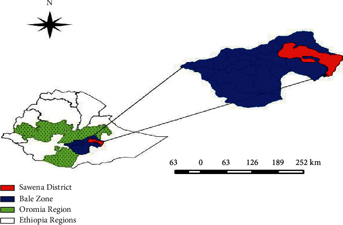
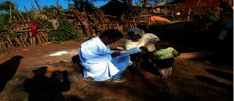
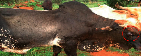
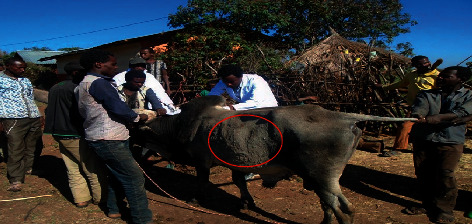
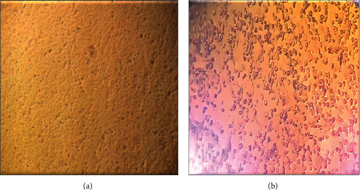
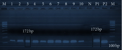
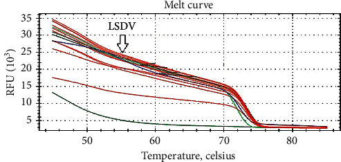
Lumpy skin disease (LSD) is a viral disease caused by LSD virus and is one of the most economically significant transboundary and emerging diseases of cattle. LSD causes considerable economic losses due to emaciation, damage to hides, infertility, and loss of milk production. In Ethiopia, the disease is distributed almost in all regions and is regarded as one of the most economically important livestock diseases in the country. An outbreak investigation of the disease was monitored from October 2016 to April 2017 in southern pastoral areas of Bale Zone, Oromia, Ethiopia. In December 2016, LSD outbreak occurred in Sawena district of Bale Zone, from which necessary biopsy samples were collected from actively infected animals for the purpose of virus isolation, and characterization using different molecular techniques at National Animal Health and Diagnostic Investigation Center (NAHDIC) of Sebeta, Ethiopia. In addition, clinical examination of infected and in-contact animals was carried out together with a questionnaire survey. Based on the clinical manifestations, LSD was recorded in 18% (94/522) of examined cattle, whereas biopsy samples from 20 clinically positive animals were collected for further laboratory process. The morbidity rate was higher in animals less than two years 28.97% (31/107) than other ages and showed a statistically significant difference with < 0.05. Female animals showed higher morbidity rate of 20.59% (76/369) than male animals (11.76%) (18/153) with a significant difference at ≤ 0.003. Mortality rate and case fatality were also significantly higher in young animals than other age groups. Viruses were isolated from both skin biopsies and nasal swabs on Vero cell line. From both skin biopsies and nasal swabs, the virus DNA was identified by amplifying the 172 bp DNA fragment using real-time and conventional PCR. Providing adequate diagnostic facilities, establishing strategic policies for effective control and eradication and awareness creations for communities for early identification or reporting were recommendations made to minimize economic losses of the disease.

摘要

结节性皮肤病(LSD)是一种由结节性皮肤病病毒引起的病毒性疾病,是牛类最具经济重要性的跨境和新出现疾病之一。结节性皮肤病由于消瘦、皮革受损、不育和产奶量下降而造成相当大的经济损失。在埃塞俄比亚,该病几乎分布于所有地区,被视为该国最具经济重要性的家畜疾病之一。2016年10月至2017年4月,在埃塞俄比亚奥罗米亚州巴勒区南部牧区对该病进行了疫情调查。2016年12月,巴勒区索韦纳区爆发了结节性皮肤病,从活跃感染动物身上采集了必要的活检样本,以便在埃塞俄比亚塞贝塔的国家动物卫生和诊断调查中心(NAHDIC)进行病毒分离,并使用不同的分子技术进行鉴定。此外,还对感染动物和接触动物进行了临床检查,并开展了问卷调查。根据临床表现,在检查的牛中,18%(94/522)被记录为患有结节性皮肤病,而从20只临床阳性动物身上采集了活检样本用于进一步的实验室检测。两岁以下动物的发病率为28.97%(31/107),高于其他年龄段,且差异具有统计学意义(<0.05)。雌性动物的发病率为20.59%(76/369),高于雄性动物(11.76%)(18/153),差异具有显著性(≤0.003)。幼龄动物的死亡率和病死率也显著高于其他年龄组。在Vero细胞系上从皮肤活检样本和鼻拭子中均分离出病毒。通过实时和常规PCR扩增172bp DNA片段,从皮肤活检样本和鼻拭子中均鉴定出病毒DNA。建议提供充足的诊断设施,制定有效的控制和根除战略政策,并提高社区的认识以实现早期识别或报告,以尽量减少该病的经济损失。

https://cdn.ncbi.nlm.nih.gov/pmc/blobs/b2e1/7929688/1d7f239e441a/VMI2021-8862180.007.jpg
https://cdn.ncbi.nlm.nih.gov/pmc/blobs/b2e1/7929688/cba15ac503e4/VMI2021-8862180.001.jpg
https://cdn.ncbi.nlm.nih.gov/pmc/blobs/b2e1/7929688/c90043f93e67/VMI2021-8862180.002.jpg
https://cdn.ncbi.nlm.nih.gov/pmc/blobs/b2e1/7929688/fce2dea1b431/VMI2021-8862180.003.jpg
https://cdn.ncbi.nlm.nih.gov/pmc/blobs/b2e1/7929688/78d4567afd5a/VMI2021-8862180.004.jpg
https://cdn.ncbi.nlm.nih.gov/pmc/blobs/b2e1/7929688/8ffac45c42b5/VMI2021-8862180.005.jpg
https://cdn.ncbi.nlm.nih.gov/pmc/blobs/b2e1/7929688/de251d08113c/VMI2021-8862180.006.jpg
https://cdn.ncbi.nlm.nih.gov/pmc/blobs/b2e1/7929688/1d7f239e441a/VMI2021-8862180.007.jpg

相似文献

1
Molecular Characterization of Lumpy Skin Disease Virus Isolates from Outbreak Cases in Cattle from Sawena District of Bale Zone, Oromia, Ethiopia.埃塞俄比亚奥罗米亚州巴勒区萨韦纳地区牛群爆发病例中结节性皮肤病病毒分离株的分子特征分析
Vet Med Int. 2021 Feb 4;2021:8862180. doi: 10.1155/2021/8862180. eCollection 2021.
2
Molecular Detection and Isolation of Lumpy Skin Disease Virus during an Outbreak in West Hararghe Zone, Eastern Ethiopia.埃塞俄比亚东部哈拉尔格西部地区疫情期间结节性皮肤病病毒的分子检测与分离
Vet Med Int. 2024 Feb 29;2024:9487970. doi: 10.1155/2024/9487970. eCollection 2024.
3
Economic impact of lumpy skin disease and cost effectiveness of vaccination for the control of outbreaks in Ethiopia.埃塞俄比亚牛结节性皮肤病的经济影响及疫苗接种控制疫情的成本效益
Prev Vet Med. 2017 Nov 1;147:100-107. doi: 10.1016/j.prevetmed.2017.09.003. Epub 2017 Sep 8.
4
Outbreak Investigation, Isolation, and Molecular Characterization of Lumpy Skin Disease Virus in Cattle from North West Oromia Region, Ethiopia.埃塞俄比亚奥罗米亚州西北部牛群中结节性皮肤病病毒的暴发调查、隔离及分子特征分析
Vet Med Int. 2024 Aug 16;2024:6038724. doi: 10.1155/2024/6038724. eCollection 2024.
5
Molecular detection and characterization of lumpy skin disease viruses from outbreaks in Thailand in 2021.2021年泰国疫情中结节性皮肤病病毒的分子检测与特征分析
Transbound Emerg Dis. 2022 Sep;69(5):e2145-e2152. doi: 10.1111/tbed.14552. Epub 2022 Apr 18.
6
Molecular identification and risk factor analysis of the first Lumpy skin disease outbreak in cattle in Mongolia.蒙古首次牛传染性块状皮肤病暴发的分子鉴定及危险因素分析。
J Vet Med Sci. 2022 Sep 5;84(9):1244-1252. doi: 10.1292/jvms.22-0250. Epub 2022 Jul 18.
7
Epidemiological Study of Lumpy Skin Disease Outbreaks in North-western Iran.伊朗西北部块状皮肤病暴发的流行病学研究。
Transbound Emerg Dis. 2017 Dec;64(6):1782-1789. doi: 10.1111/tbed.12565. Epub 2016 Sep 15.
8
Lumpy skin disease in cattle in central Ethiopia: outbreak investigation and isolation and molecular detection of the virus.埃塞俄比亚中部牛群的结节性皮肤病:疫情调查、病毒分离与分子检测
Rev Sci Tech. 2014 Dec;33(3):877-87. doi: 10.20506/rst.33.3.2325.
9
Spatiotemporal analysis and forecasting of lumpy skin disease outbreaks in Ethiopia based on retrospective outbreak reports.基于回顾性疫情报告的埃塞俄比亚牛结节性皮肤病疫情时空分析与预测
Front Vet Sci. 2024 Mar 12;11:1277007. doi: 10.3389/fvets.2024.1277007. eCollection 2024.
10
Observations on presumptive lumpy skin disease in native cattle and Asian water buffaloes around the tiger reserves of the central Indian highlands.对中央印度高原老虎保护区周围本地黄牛和亚洲水牛疑似牛结节疹病的观察。
N Z Vet J. 2022 Mar;70(2):101-108. doi: 10.1080/00480169.2021.1984335. Epub 2021 Oct 25.

引用本文的文献

1
Prevalence of Lumpy Skin Disease in Africa: A Systematic Review and Meta-Analysis from 2007 to 2023.非洲牛结节性皮肤病的患病率:2007年至2023年的系统评价和荟萃分析
Vet Med Int. 2024 Jun 5;2024:9991106. doi: 10.1155/2024/9991106. eCollection 2024.
2
In-silico characterization of LSDV132 protein divulged its BCL-2-like nature.绵羊痘病毒132蛋白的计算机模拟特征揭示了其类BCL-2的性质。
Heliyon. 2024 Mar 11;10(6):e27657. doi: 10.1016/j.heliyon.2024.e27657. eCollection 2024 Mar 30.
3
Molecular Detection and Isolation of Lumpy Skin Disease Virus during an Outbreak in West Hararghe Zone, Eastern Ethiopia.

本文引用的文献

1
Lumpy skin disease in cattle in central Ethiopia: outbreak investigation and isolation and molecular detection of the virus.埃塞俄比亚中部牛群的结节性皮肤病:疫情调查、病毒分离与分子检测
Rev Sci Tech. 2014 Dec;33(3):877-87. doi: 10.20506/rst.33.3.2325.
2
Development of a cost-effective method for capripoxvirus genotyping using snapback primer and dsDNA intercalating dye.利用回套引物和双链 DNA 嵌入染料开发一种具有成本效益的羊痘病毒基因分型方法。
PLoS One. 2013 Oct 7;8(10):e75971. doi: 10.1371/journal.pone.0075971. eCollection 2013.
3
Risk assessments of lumpy skin diseases in Borena bull market chain and its implication for livelihoods and international trade.
埃塞俄比亚东部哈拉尔格西部地区疫情期间结节性皮肤病病毒的分子检测与分离
Vet Med Int. 2024 Feb 29;2024:9487970. doi: 10.1155/2024/9487970. eCollection 2024.
4
Isolation, adaptation, and characterization of lumpy skin disease virus from cattle in Bangladesh.从孟加拉国牛群中分离、适应和鉴定结节性皮肤病病毒
J Adv Vet Anim Res. 2023 Sep 30;10(3):563-569. doi: 10.5455/javar.2023.j710. eCollection 2023 Sep.
5
Lumpy skin disease: A newly emerging disease in Southeast Asia.结节性皮肤病:东南亚一种新出现的疾病。
Vet World. 2022 Dec;15(12):2764-2771. doi: 10.14202/vetworld.2022.2764-2771. Epub 2022 Dec 5.
6
Insights into the Prognostic Role of Serum Interleukin-6 and Hematobiochemical Alterations in Cattle during Recent Outbreaks of Lumpy Skin Disease in Lodhran District, Pakistan.巴基斯坦洛德兰地区近期牛结节性皮肤病疫情期间血清白细胞介素-6及血液生化改变的预后作用洞察
Vaccines (Basel). 2023 Jan 3;11(1):113. doi: 10.3390/vaccines11010113.
7
Molecular diagnosis of three outbreaks during three successive years (2018, 2019, and 2020) of in cattle in Sharkia Governorate, Egypt.埃及 Sharkia 省 2018、2019 和 2020 连续三年牛群暴发疫情的分子诊断。
Open Vet J. 2022 Jul-Aug;12(4):451-462. doi: 10.5455/OVJ.2022.v12.i4.6. Epub 2022 Jul 12.
8
Molecular identification and characterization of Lumpy skin disease virus emergence from cattle in the northeastern part of Thailand.从泰国东北部牛中出现的块状皮肤病病毒的分子鉴定和特征描述。
J Vet Sci. 2022 Sep;23(5):e73. doi: 10.4142/jvs.22111. Epub 2022 Aug 9.
9
Clinicopathological and molecular studies on cattle naturally infected with lumpy skin diseases in selected districts of Wolaita Zone, Southern Ethiopia.在埃塞俄比亚南部沃莱塔地区选定的地区,对自然感染块状皮肤病的牛进行临床病理和分子研究。
BMC Vet Res. 2022 Aug 3;18(1):297. doi: 10.1186/s12917-022-03403-4.
10
First molecular characterization of poxviruses in cattle, sheep, and goats in Botswana.博茨瓦纳牛、绵羊和山羊痘病毒的首次分子特征描述。
Virol J. 2021 Aug 14;18(1):167. doi: 10.1186/s12985-021-01634-9.
博雷纳牛市链中牛结节性皮肤病的风险评估及其对生计和国际贸易的影响。
Trop Anim Health Prod. 2013 Jun;45(5):1153-9. doi: 10.1007/s11250-012-0340-9. Epub 2012 Dec 29.
4
Review: lumpy skin disease: an emerging threat to Europe, the Middle East and Asia.综述:块状皮肤病:对欧洲、中东和亚洲的新威胁。
Transbound Emerg Dis. 2012 Feb;59(1):40-8. doi: 10.1111/j.1865-1682.2011.01242.x. Epub 2011 Jul 12.
5
Use of the Capripoxvirus homologue of Vaccinia virus 30 kDa RNA polymerase subunit (RPO30) gene as a novel diagnostic and genotyping target: development of a classical PCR method to differentiate Goat poxvirus from Sheep poxvirus.利用羊痘病毒与牛痘病毒 30kDa RNA 聚合酶亚基(RPO30)基因的同源物作为一种新的诊断和基因分型靶标:建立一种区分山羊痘病毒和绵羊痘病毒的经典 PCR 方法。
Vet Microbiol. 2011 Apr 21;149(1-2):30-9. doi: 10.1016/j.vetmic.2010.09.038. Epub 2010 Oct 17.
6
Risk factors associated with observed clinical lumpy skin disease in Ethiopia.与埃塞俄比亚观察到的临床块状皮肤病相关的风险因素。
Epidemiol Infect. 2010 Nov;138(11):1657-66. doi: 10.1017/S0950268810000506. Epub 2010 Mar 17.
7
The distribution of ixodid ticks (Acari: Ixodidae) in central Ethiopia.埃塞俄比亚中部硬蜱(蜱螨目:硬蜱科)的分布情况
Onderstepoort J Vet Res. 2001 Dec;68(4):243-51.
8
Sheep poxvirus identification from clinical specimens by PCR, cell culture, immunofluorescence and agar gel immunoprecipitation assay.通过聚合酶链式反应(PCR)、细胞培养、免疫荧光和琼脂凝胶免疫沉淀试验从临床标本中鉴定羊痘病毒。
Mol Cell Probes. 2000 Oct;14(5):305-10. doi: 10.1006/mcpr.2000.0319.
9
Case-cohort analysis of case-coverage studies of vaccine effectiveness.疫苗效力病例覆盖研究的病例队列分析。
Am J Epidemiol. 1995 Nov 1;142(9):1000-6. doi: 10.1093/oxfordjournals.aje.a117730.
10
Lumpy skin disease--a review.结节性皮肤病——综述
Trop Anim Health Prod. 1988 Feb;20(1):11-7. doi: 10.1007/BF02239636.