Wang Fanglan, Hujjaree Khamlesh, Wang Xiaoping
Department of Psychiatry, National Clinical Research Center for Mental Disorders, The Second Xiangya Hospital of Central South University, Changsha, China.
Front Psychiatry. 2021 Feb 26;12:638722. doi: 10.3389/fpsyt.2021.638722. eCollection 2021.
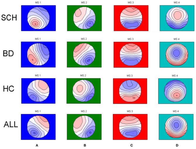
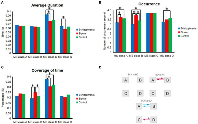
Schizophrenia (SCH) and bipolar disorder (BD) are characterized by many types of symptoms, damaged cognitive function, and abnormal brain connections. The microstates are considered to be the cornerstones of the mental states shown in EEG data. In our study, we investigated the use of microstates as biomarkers to distinguish patients with bipolar disorder from those with schizophrenia by analyzing EEG data measured in an eyes-closed resting state. The purpose of this article is to provide an electron directional physiological explanation for the observed brain dysfunction of schizophrenia and bipolar disorder patients. We used microstate resting EEG data to explore group differences in the duration, coverage, occurrence, and transition probability of 4 microstate maps among 20 SCH patients, 26 BD patients, and 35 healthy controls (HCs). Microstate analysis revealed 4 microstates (A-D) in global clustering across SCH patients, BD patients, and HCs. The samples were chosen to be matched. We found the greater presence of microstate B in BD patients, and the less presence of microstate class A and B, the greater presence of microstate class C, and less presence of D in SCH patients. Besides, a greater frequent switching between microstates A and B and between microstates B and A in BD patients than in SCH patients and HCs and less frequent switching between microstates C and D and between microstates D and C in BD patients compared with SCH patients. We found abnormal features of microstate A, B in BD patients and abnormal features of microstate A, B, C, and D in SCH patients. These features may indicate the potential abnormalities of SCH patients and BD patients in distributing neural resources and influencing opportune transitions between different states of activity.
精神分裂症(SCH)和双相情感障碍(BD)具有多种症状、受损的认知功能以及异常的脑连接。微状态被认为是脑电图(EEG)数据中所显示精神状态的基石。在我们的研究中,我们通过分析闭眼静息状态下测量的EEG数据,研究了将微状态用作生物标志物以区分双相情感障碍患者和精神分裂症患者的用途。本文的目的是为精神分裂症和双相情感障碍患者所观察到的脑功能障碍提供一种电子定向生理学解释。我们使用微状态静息EEG数据来探索20名精神分裂症患者、26名双相情感障碍患者和35名健康对照者(HCs)之间4种微状态图在持续时间、覆盖范围、出现频率和转换概率方面的组间差异。微状态分析在精神分裂症患者、双相情感障碍患者和健康对照者的全局聚类中揭示了4种微状态(A - D)。样本经过匹配选取。我们发现双相情感障碍患者中微状态B的出现频率更高,而精神分裂症患者中微状态A和B的出现频率更低、微状态C的出现频率更高以及微状态D的出现频率更低。此外,与精神分裂症患者和健康对照者相比,双相情感障碍患者中微状态A和B之间以及微状态B和A之间的转换频率更高,而双相情感障碍患者中微状态C和D之间以及微状态D和C之间的转换频率低于精神分裂症患者。我们发现双相情感障碍患者中微状态A、B存在异常特征,精神分裂症患者中微状态A、B、C和D存在异常特征。这些特征可能表明精神分裂症患者和双相情感障碍患者在分配神经资源以及影响不同活动状态之间适时转换方面存在潜在异常。