Department of Respiratory Diseases, Univ Montpellier, CHU Montpellier, Montpellier, France.
PhyMedExp, Univ Montpellier, CNRS, INSERM, CHU Montpellier, Montpellier, France.
Front Immunol. 2021 Feb 26;12:630096. doi: 10.3389/fimmu.2021.630096. eCollection 2021.
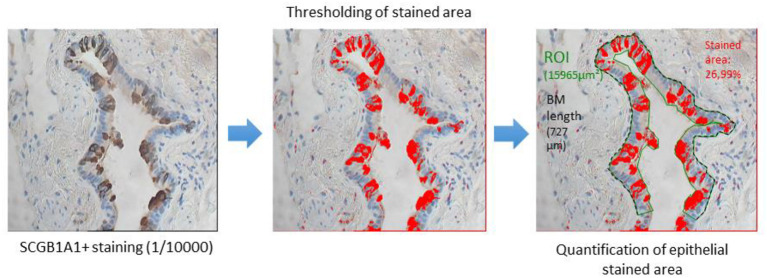
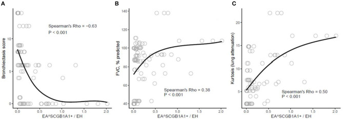
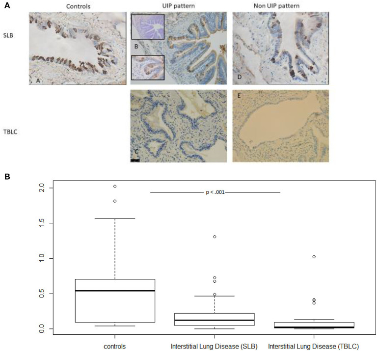
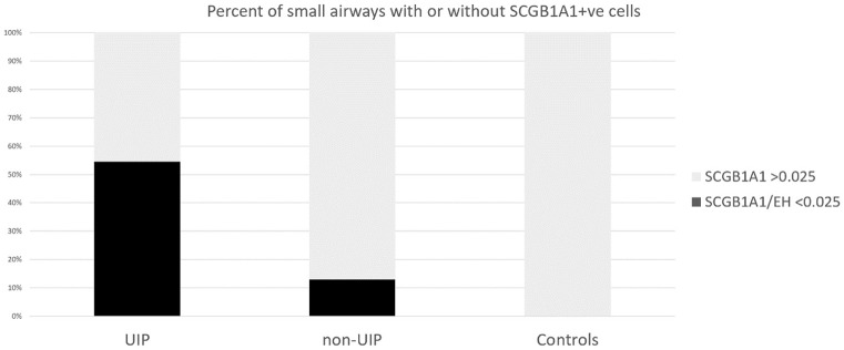
Distal airway metaplasia may precede honeycombing in progressive fibrosing interstitial lung disease (ILD). The SCGB1A1 bronchiolar-specific club cell may play a role in this aberrant regenerative process. To assess the presence of club cells in the small airways of patients suffering from ILD. Small airways (internal diameter <2 mm) in lung samples [surgical lung biopsy (SLB) and/or transbronchial lung cryobiopsy (TBLC)] from 14 patients suffering from ILD and 10 controls were morphologically assessed and stained for SCGB1A1. SCGB1A1 was weighted by epithelial height as a marker of airway generation (SCGB1A1/EH). Correlations between clinical, functional, and high-resolution CT (HRCT) prognostic factors and histomorphometry were assessed. Small airways from samples with ILD patterns were significantly less dense in terms of SCGB1A1 cells [0.064 (0.020-0.172)] as compared to controls' sample's small airways [0.393 (0.082-0.698), < 0.0001]. Usual interstitial pneumonia (UIP) patterns most frequently contained small airways with limited or absent SCGB1A1 expression (SCGB1A1/EH <0.025): UIP (18/33; 55%) as compared with non-UIP patterns (4/31; 13%) or controls (0/29; 0%): < 0.0001. In addition, correlations with HRCT indicated a significant negative relationship between SCGB1A1 and bronchiectasis as a feature of bronchiolization (Rho -0.63, < 0.001) and a positive relationship with both forced vital capacity (FVC) and Hounsfield unit (HU)-distribution pattern in kurtosis (Rho 0.38 and 0.50, respectively, both < 0.001) as markers of fibrotic changes. Compared with controls, the small airways of patients with ILD more often lack SCGB1A1, especially so in UIP. Low densities of SCGB1A1-marked cells correlate with bronchiectasis and fibrotic changes. Further research investigating SCGB1A1 staining as a pathological feature of the bronchiolization process is merited.
远端气道化生可能先于进行性纤维化间质性肺病 (ILD) 的蜂窝肺形成。SCGB1A1 支气管特异性克拉细胞可能在这一异常再生过程中发挥作用。评估患有 ILD 的患者的小气道中是否存在克拉细胞。对 14 名患有 ILD 和 10 名对照者的肺组织标本[外科肺活检 (SLB) 和/或经支气管肺冷冻活检 (TBLC)]的小气道进行形态学评估,并对 SCGB1A1 进行染色。SCGB1A1 作为气道生成的标志物(SCGB1A1/EH),按上皮高度加权。评估临床、功能和高分辨率 CT(HRCT)预后因素与组织形态计量学之间的相关性。ILD 模式样本的小气道中 SCGB1A1 细胞的密度明显较低[0.064(0.020-0.172)],而对照组样本的小气道中 SCGB1A1 细胞的密度较高[0.393(0.082-0.698)],<0.0001。常见间质性肺炎 (UIP) 模式最常包含 SCGB1A1 表达有限或缺失的小气道(SCGB1A1/EH <0.025):UIP(18/33;55%)与非 UIP 模式(4/31;13%)或对照组(0/29;0%)相比:<0.0001。此外,与 HRCT 的相关性表明,SCGB1A1 与支气管扩张呈显著负相关,支气管扩张是小气道化生的特征(Rho -0.63,<0.001),与用力肺活量 (FVC) 和峰度 HU 分布模式呈正相关(Rho 0.38 和 0.50,均<0.001),这是纤维化变化的标志物。与对照组相比,ILD 患者的小气道中更常缺乏 SCGB1A1,尤其是 UIP 患者。SCGB1A1 标记细胞的密度较低与支气管扩张和纤维化变化相关。进一步研究 SCGB1A1 染色作为小气道化生过程的病理特征是值得的。