Williams James M, Adamo Daniel A, Olson Michael C
Department of Radiology, Mayo Clinic College of Medicine, Mayo Clinic, 200 First Street SW, Rochester, MN, 55905 USA.
Radiol Case Rep. 2021 Feb 24;16(5):1072-1074. doi: 10.1016/j.radcr.2021.02.034. eCollection 2021 May.
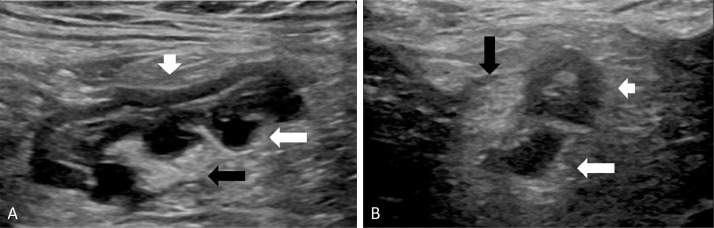
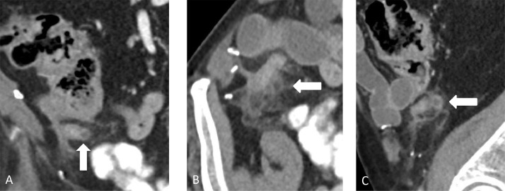
Acute appendiceal diverticulitis is a difficult prospective diagnosis due to both its rarity and clinical overlap with other ileocecal diseases, such as acute appendicitis. Few preoperative radiological images of acute appendiceal diverticulitis have been presented in the literature. We report a case of acute appendiceal diverticulitis initially diagnosed with outpatient ultrasound and confirmed with contrast-enhanced computed tomography in an 85-year-old male who presented with right lower quadrant pain. Appendiceal diverticulitis is an often overlooked diagnosis, and given its association with appendiceal neoplasms and relatively high rate of perforation, it is important to consider this entity in patients with right lower quadrant pain in order to ensure that they are managed appropriately. The addition of high quality ultrasound and computed tomography (CT) images to the literature may heighten awareness of this rare condition and facilitate accurate preoperative diagnosis and management in these patients.
急性阑尾憩室炎由于其罕见性以及与其他回盲部疾病(如急性阑尾炎)在临床上的重叠,很难进行前瞻性诊断。文献中很少有急性阑尾憩室炎的术前影像学图像。我们报告一例85岁男性患者,最初通过门诊超声诊断为急性阑尾憩室炎,并经增强计算机断层扫描确诊,该患者表现为右下腹疼痛。阑尾憩室炎是一种常被忽视的诊断,鉴于其与阑尾肿瘤的关联以及相对较高的穿孔率,对于右下腹疼痛的患者考虑这一病症很重要,以便确保他们得到适当的治疗。高质量的超声和计算机断层扫描(CT)图像补充到文献中,可能会提高对这种罕见病症的认识,并有助于这些患者进行准确的术前诊断和治疗。