Rifai Olivia M, McGrory Sarah, Robbins Cason B, Grewal Dilraj S, Liu Andy, Fekrat Sharon, MacGillivray Thomas J
Translational Neuroscience PhD Programme University of Edinburgh Edinburgh UK.
Centre for Clinical Brain Sciences College of Medicine and Veterinary Medicine University of Edinburgh Edinburgh UK.
Alzheimers Dement (Amst). 2021 Mar 3;13(1):e12149. doi: 10.1002/dad2.12149. eCollection 2021.
Discovering non-invasive and easily acquired biomarkers that are conducive to the accurate diagnosis of dementia is an urgent area of ongoing clinical research. One promising approach is retinal imaging, as there is homology between retinal and cerebral vasculature. Recently, optical coherence tomography angiography (OCT-A) has emerged as a promising new technology for imaging the microvasculature of the retina.
A systematic review and meta-analysis was conducted to examine the application of OCT-A in dementia.
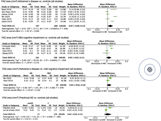
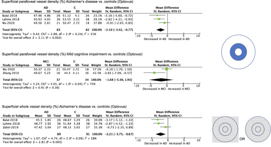
Fourteen studies assessing OCT-A in preclinical Alzheimer's disease (AD), mild cognitive impairment, or AD were included. Exploratory meta-analyses revealed a significant increase in the foveal avascular zone area and a significant decrease in superficial parafoveal and whole vessel density in AD, although there was significant heterogeneity between studies.
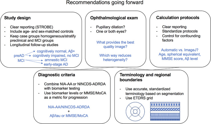
Although certain OCT-A metrics may have the potential to serve as biomarkers for AD, the field requires further standardization to allow conclusions to be reached regarding their clinical utility.
发现有助于痴呆症准确诊断的非侵入性且易于获取的生物标志物是当前临床研究的一个紧迫领域。一种有前景的方法是视网膜成像,因为视网膜和脑血管系统存在同源性。最近,光学相干断层扫描血管造影(OCT - A)已成为一种用于视网膜微血管成像的有前景的新技术。
进行了一项系统综述和荟萃分析,以研究OCT - A在痴呆症中的应用。
纳入了14项评估OCT - A在临床前阿尔茨海默病(AD)、轻度认知障碍或AD中的研究。探索性荟萃分析显示,AD患者的中心凹无血管区面积显著增加,而中心凹旁浅表和全血管密度显著降低,尽管各研究之间存在显著异质性。
尽管某些OCT - A指标可能有潜力作为AD的生物标志物,但该领域需要进一步标准化,以便就其临床效用得出结论。