Radiology, Mayo Clinic, Rochester, Minnesota, USA.
Neurosurgery, Mayo Clinic, Rochester, Minnesota, USA.
J Neurointerv Surg. 2021 Sep;13(9):816-822. doi: 10.1136/neurintsurg-2020-017133. Epub 2021 Mar 15.
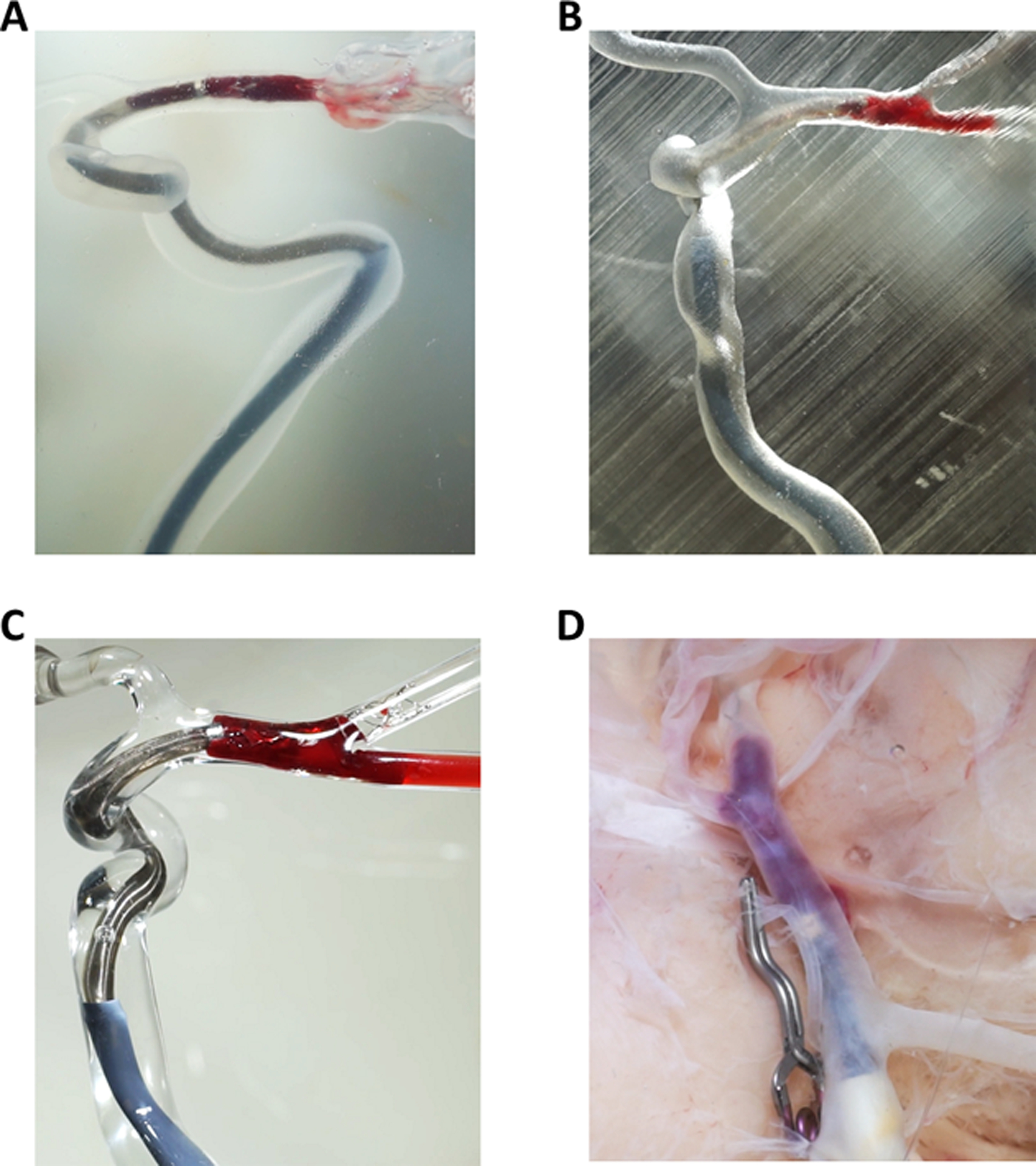
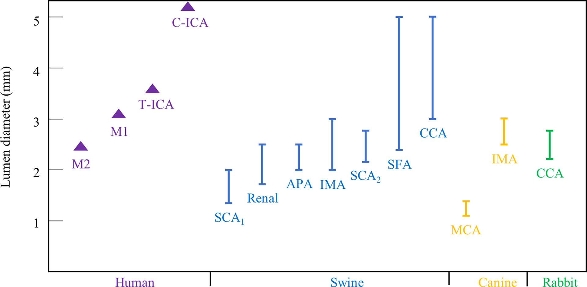
Preclinical testing platforms have been instrumental in the research and development of thrombectomy devices. However, there is no single model which fully captures the complexity of cerebrovascular anatomy, physiology, and the dynamic artery-clot-device interaction. This article provides a critical review of phantoms, in-vivo animal, and human cadaveric models used for thrombectomy testing and provides insights into the strengths and limitations of each platform. Articles published in the past 10 years that reported thrombectomy testing platforms were identified. Characteristics of each test platform, such as intracranial anatomy, artery tortuosity, vessel friction, flow conditions, device-vessel interaction, and visualization, were captured and benchmarked against human cerebral vessels involved in large-vessel occlusion stroke. Thrombectomy phantoms have been constructed from silicone, direct 3D-printed polymers, and glass. These phantoms represent oversimplified patient-specific cerebrovascular geometry but enable adequate visualization of devices and clots under appropriate flow conditions. They do not realistically mimic the artery-clot interaction. For the animal models, arteries from swine, canines, and rabbits have been reported. These models can reasonably replicate the artery-clot-device interaction and have the unique value of evaluating the safety of thrombectomy devices. However, the vasculature geometries are substantially less complex and flow conditions are different from human cerebral arteries. Cadaveric models are the most accurate vascular representations but with limited access and challenges in reproducibility of testing conditions. Multiple test platforms should be likely used for comprehensive evaluation of thrombectomy devices. Interpretation of the testing results should take into consideration platform-specific limitations.
临床前测试平台在血栓切除术设备的研究和开发中发挥了重要作用。然而,没有一种单一的模型能够完全捕捉到脑血管解剖、生理学以及动态动脉-血栓-器械相互作用的复杂性。本文对用于血栓切除术测试的模型、体内动物和人体尸体模型进行了批判性评价,并深入探讨了每个平台的优缺点。本文回顾了过去 10 年中发表的报道血栓切除术测试平台的文章。对每个测试平台的特征进行了描述,如颅内解剖结构、动脉迂曲度、血管摩擦力、流动条件、器械-血管相互作用以及可视化程度,并与涉及大血管闭塞性中风的人类大脑血管进行了对比。血栓切除术模型由硅酮、直接 3D 打印聚合物和玻璃制成。这些模型虽然代表了过于简化的患者特定的脑血管几何形状,但能够在适当的流动条件下充分观察器械和血栓。然而,这些模型无法真实模拟动脉-血栓相互作用。对于动物模型,已经报道了来自猪、犬和兔的动脉。这些模型可以合理地模拟动脉-血栓-器械相互作用,具有评估血栓切除术器械安全性的独特价值。然而,这些模型的血管几何形状要简单得多,流动条件也与人类大脑动脉不同。尸体模型是最准确的血管模型,但由于获取途径有限,以及测试条件的重现性存在挑战,因此其应用受到限制。为了全面评估血栓切除术器械,可能需要使用多种测试平台。在解释测试结果时,应考虑到平台特定的局限性。