Department of Radiology, Charité-Universitätsmedizin Berlin, Berlin, Germany.
Department of Radiology, Vivantes Klinikum am Friedrichshain, Berlin, Germany.
Sci Rep. 2021 Mar 15;11(1):5903. doi: 10.1038/s41598-021-85303-5.
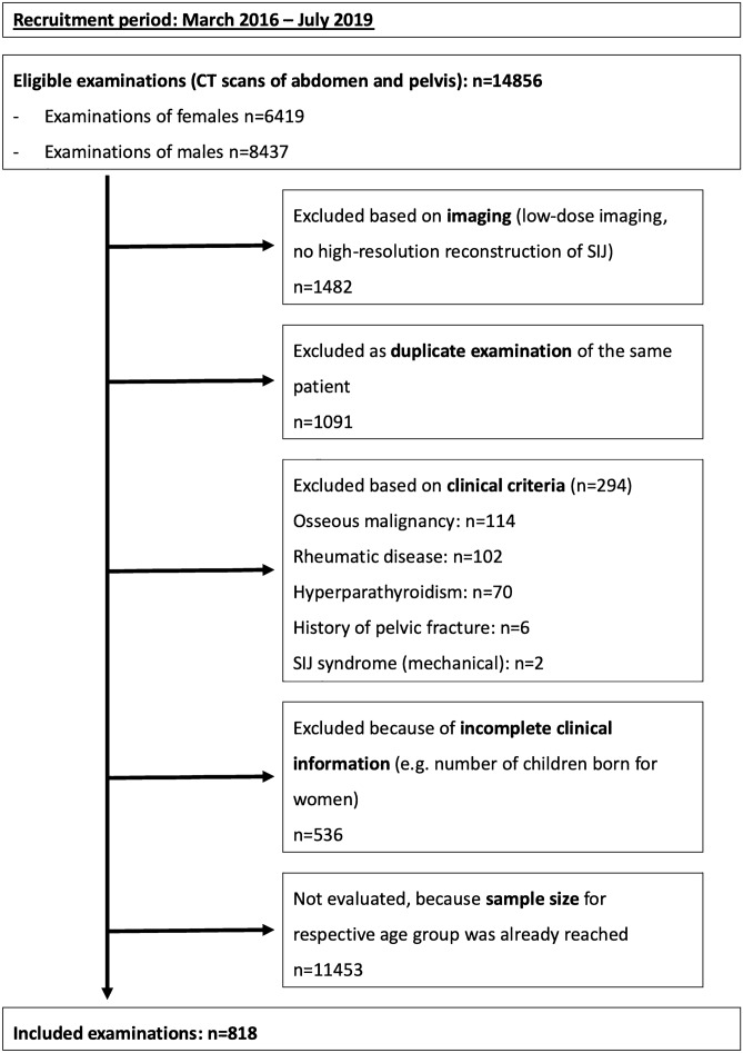
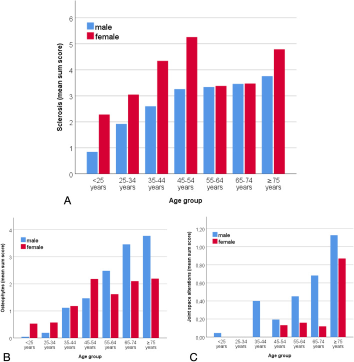
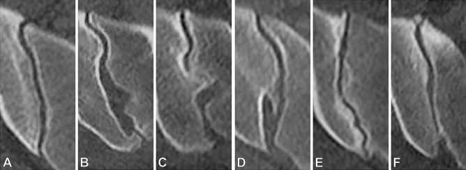
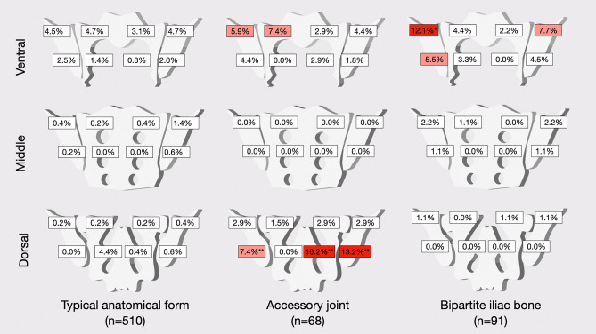
Degeneration of the sacroiliac joints (SIJs) is a common finding, while its underlying cause and development remain incompletely understood. The aim of this investigation was to describe the spatial distribution of degenerative SIJ changes across age groups and to investigate for the first time their relationship to anatomical form and sex. For this IRB-approved investigation, demographic data of 818 patients without SIJ disease were retrieved from electronic patient records. High-resolution computed tomography (CT) datasets of all patients were analysed retrospectively for seven predefined age groups (ten-year increments, from < 25 to ≥ 75). A structured scoring system was applied to assess sclerosis, osteophytes, joint space alterations, and anatomical form. Chi-square tests were used to compare frequencies of degenerative lesions, and logistic regression analyses were performed to investigate associations between demographic data, anatomical form, and the presence of structural lesions. Sclerosis and osteophytes were common findings, with an overall prevalence of 45.7% and 46.8%, respectively. Female sex had an odds ratio (OR) of 0.15 (95% CI: 0.08-0.27) for the presence of ventral osteophytes and of 4.42 (95% CI: 2.77-7.04) for dorsal osteophytes. Atypical joint forms were significantly more prevalent in women with 62.1% vs. 14.1% in men (p < 0.001). Accessory joints increased the likelihood of dorsal sclerosis (OR 2.735; 95% CI 1.376-5.436) while a typical joint form decreased its likelihood (OR 0.174; 95% CI 0.104-0.293). Sex and anatomical joint form have a major impact on the development of degenerative lesions of the SIJs and their spatial distribution.
骶髂关节(SIJ)退变是一种常见的表现,但其潜在病因和发展仍不完全清楚。本研究旨在描述不同年龄组 SIJ 退变的空间分布,并首次探讨其与解剖形态和性别的关系。这项经过机构审查委员会批准的研究中,从电子病历中检索了 818 名无 SIJ 疾病的患者的人口统计学数据。回顾性分析了所有患者的高分辨率计算机断层扫描(CT)数据集,分为七个预设年龄组(每 10 年为一个组,从<25 岁到≥75 岁)。应用结构化评分系统评估硬化、骨赘、关节间隙改变和解剖形态。采用卡方检验比较退行性病变的发生率,采用逻辑回归分析人口统计学数据、解剖形态与结构病变之间的关系。硬化和骨赘是常见的发现,总体患病率分别为 45.7%和 46.8%。女性发生腹侧骨赘的优势比(OR)为 0.15(95%CI:0.08-0.27),发生背侧骨赘的 OR 为 4.42(95%CI:2.77-7.04)。异常关节形态在女性中更为常见(62.1% vs. 14.1%,p<0.001)。副关节增加了背侧硬化的可能性(OR 2.735;95%CI 1.376-5.436),而典型关节形态降低了其可能性(OR 0.174;95%CI 0.104-0.293)。性别和解剖关节形态对 SIJ 退行性病变的发展及其空间分布有重大影响。