Muellner Maximilian, Kreutzinger Virginie, Becker Luis, Diekhoff Torsten, Pumberger Matthias, Schömig Friederike, Heyland Mark, Ziegeler Katharina
Center for Musculoskeletal Surgery, Charité-University Medicine Berlin, 10117 Berlin, Germany.
Department of Radiology, Vivantes Klinikum im Friedrichshain, 10249 Berlin, Germany.
Diagnostics (Basel). 2022 Jan 21;12(2):275. doi: 10.3390/diagnostics12020275.
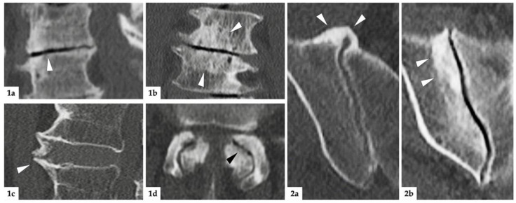
The relationship between degenerative changes of the sacroiliac joints and the lumbar spine on CT has not been studied yet. The aim of this analysis is to determine the nature of their association as well as the influence of fixed anatomical spinopelvic parameters on sacroiliac joint degeneration. For this institutional review-board-approved investigation, imaging datasets as well as electronic medical records of 719 patients without back pain from the clinical routine of our department of radiology were included. Age, sex, weight category (slim, normal, obese), parity in women and indication for imaging were noted for all patients. The presence of degenerative lesions of the lumbar spine (disc degeneration, endplate degeneration, spondylophytes, and facet joint osteoarthritis) was noted separately at each lumbar segment (L1 to L5). Sacroiliac joints were assessed for sclerosis and osteophytes. Fixed anatomical spinopelvic parameters were measured: pelvic radius = PR; pelvic incidence = PI; sacral table angle = STA. Correlation as well as regression analyses were performed; data were analyzed for males and females separately. PI increased significantly with age in both women and men, while STA decreased and PR remained constant; neither of them was associated with SIJ degeneration. SIJ degeneration correlated with disc degeneration (tau = 0.331; < 0.001), spondylophytes (tau = 0.397; < 0.001), and facet joint degeneration (tau = 0.310; < 0.001) in men, but with no parameter of spinal degeneration in women. Lumbar spinal degeneration increased the risk of sacroiliac joint degeneration in men significantly (OR 7.2; 95%CI 2.8-19.0), but it was not a significant covariable in women. Fixed spinopelvic parameters have little impact on sacroiliac joint degeneration. The degeneration of the sacroiliac joints and the lumbar spine appear to be parallel processes in men, but are largely unrelated in women.
骶髂关节与腰椎退变在CT上的关系尚未得到研究。本分析的目的是确定它们之间关联的性质以及固定的解剖学脊柱骨盆参数对骶髂关节退变的影响。对于这项经机构审查委员会批准的调查,纳入了来自我们放射科临床常规的719例无背痛患者的影像数据集以及电子病历。记录了所有患者的年龄、性别、体重类别(消瘦、正常、肥胖)、女性的妊娠次数以及成像指征。分别在每个腰椎节段(L1至L5)记录腰椎退变病变(椎间盘退变、终板退变、骨赘和小关节骨关节炎)的存在情况。评估骶髂关节的硬化和骨赘情况。测量固定的解剖学脊柱骨盆参数:骨盆半径=PR;骨盆倾斜角=PI;骶骨平台角=STA。进行了相关性和回归分析;分别对男性和女性的数据进行了分析。PI在女性和男性中均随年龄显著增加,而STA降低,PR保持不变;它们均与骶髂关节退变无关。在男性中,骶髂关节退变与椎间盘退变(tau=0.331;P<0.001)、骨赘(tau=0.397;P<0.001)和小关节退变(tau=0.310;P<0.001)相关,但在女性中与任何脊柱退变参数均无关。腰椎退变显著增加了男性骶髂关节退变的风险(OR 7.2;95%CI 2.8 - 19.0),但在女性中不是一个显著的协变量。固定的脊柱骨盆参数对骶髂关节退变影响很小。骶髂关节和腰椎的退变在男性中似乎是平行的过程,但在女性中基本无关。