Department of Cariology, Restorative Sciences and Endodontics, University of Michigan School of Dentistry, 1011 N. University Rm. G049, Ann Arbor, MI 48109-1078,
Eur Cell Mater. 2021 Mar 16;41:332-344. doi: 10.22203/eCM.v041a21.
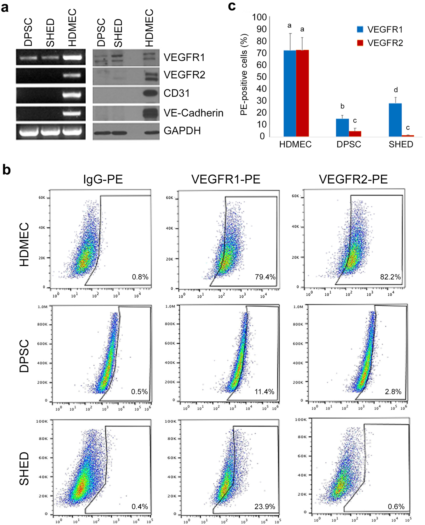
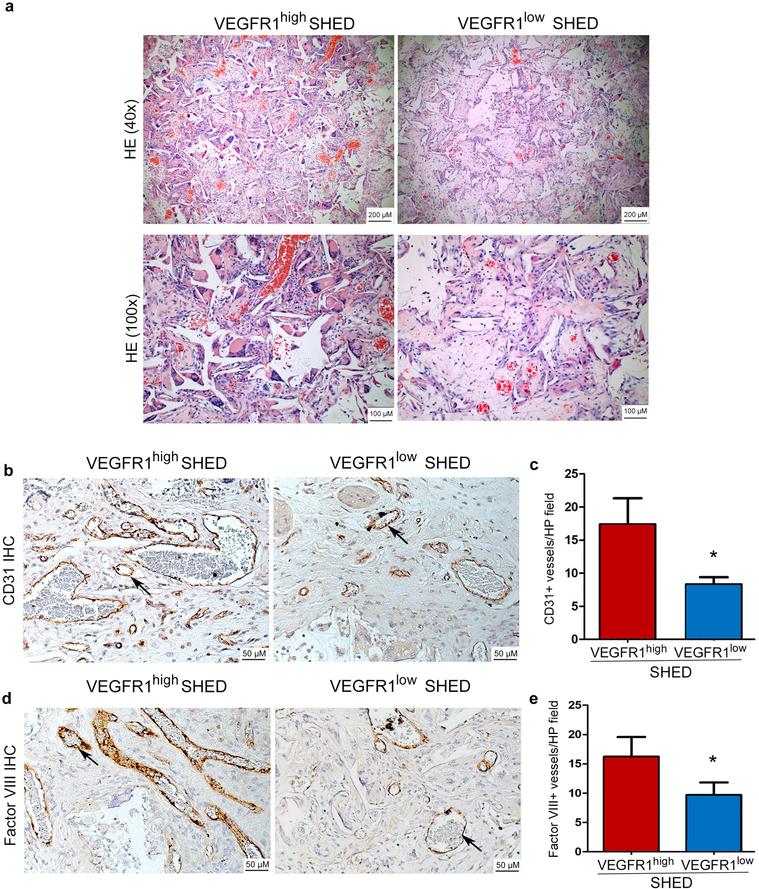
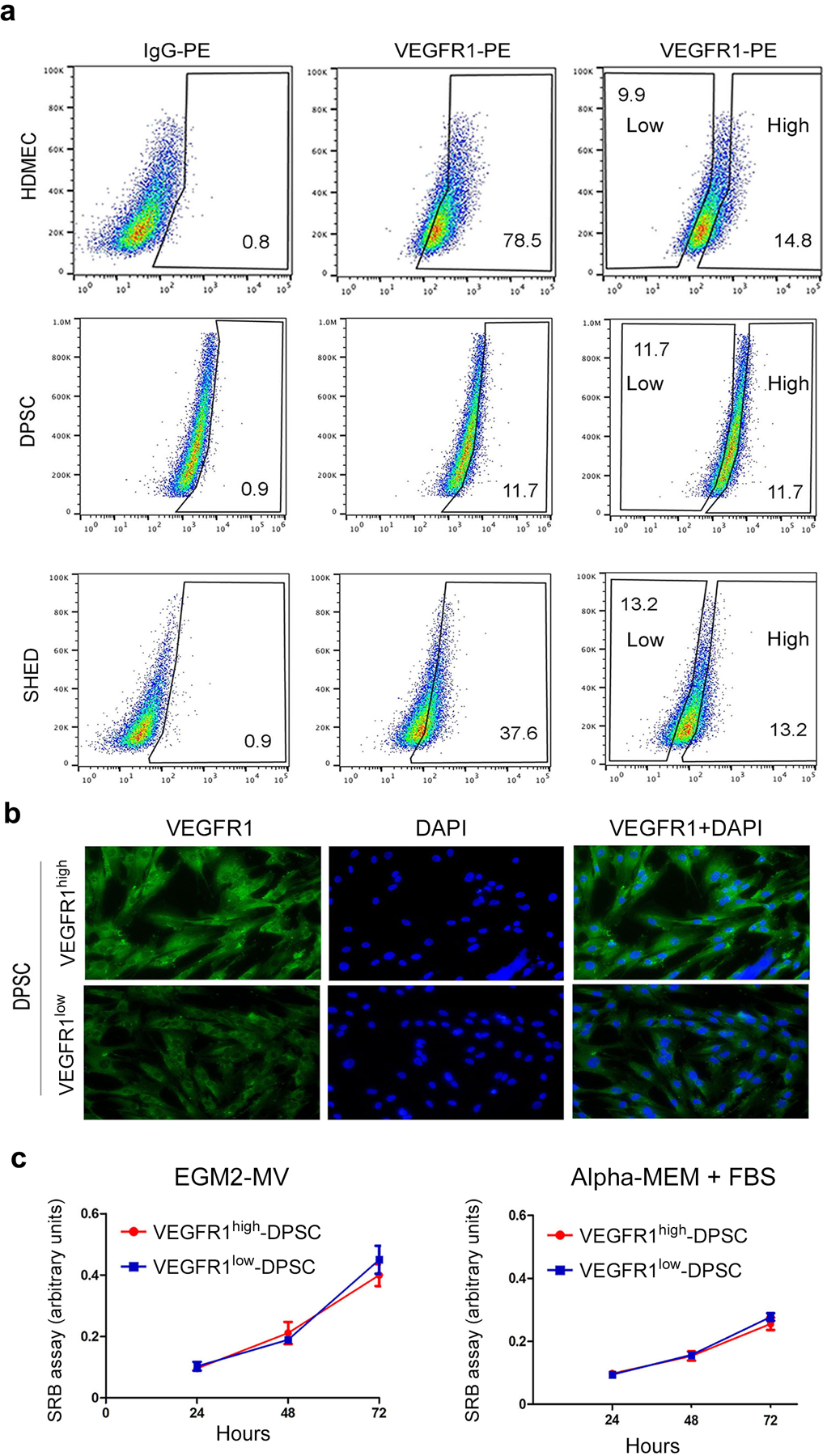
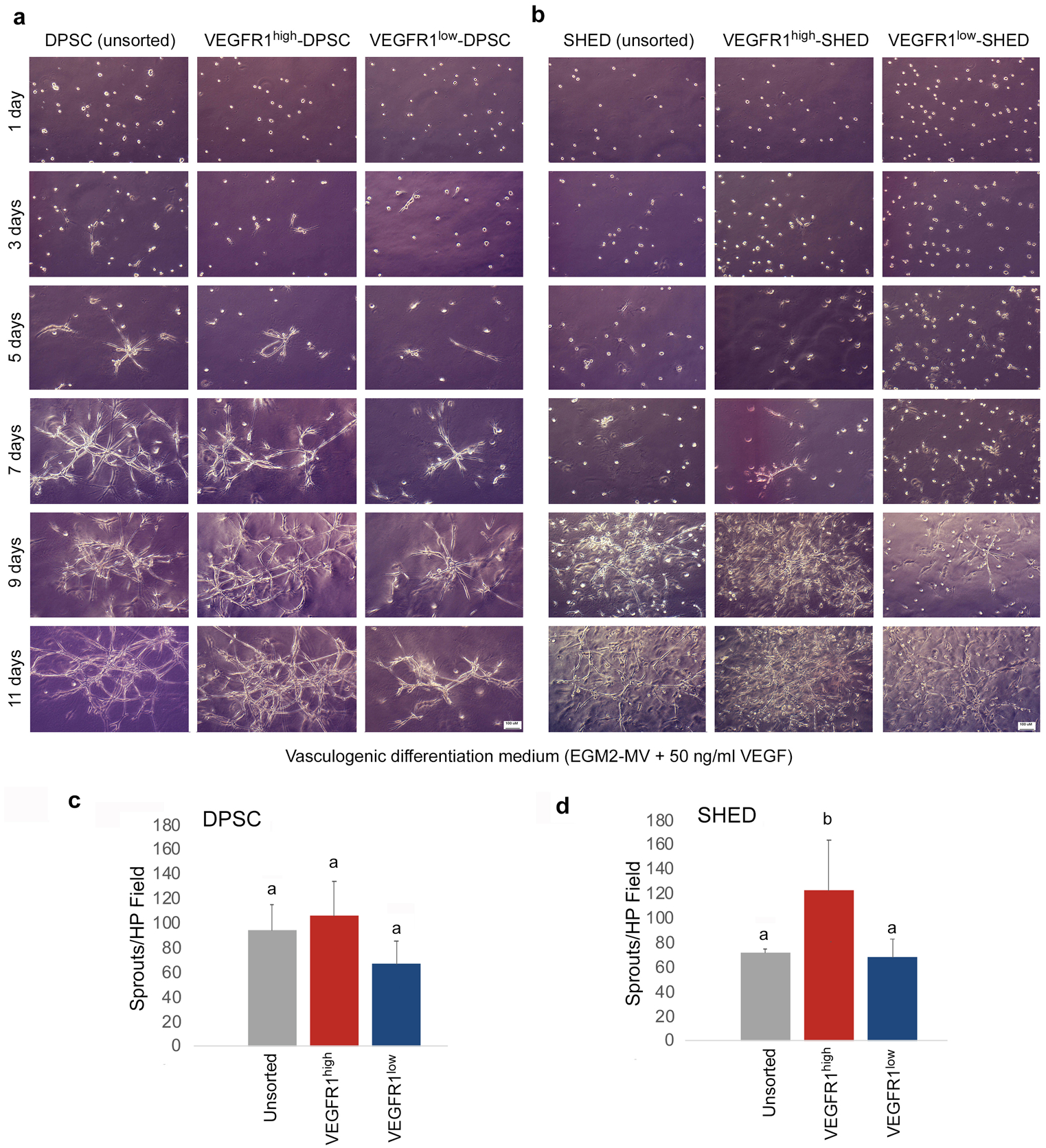
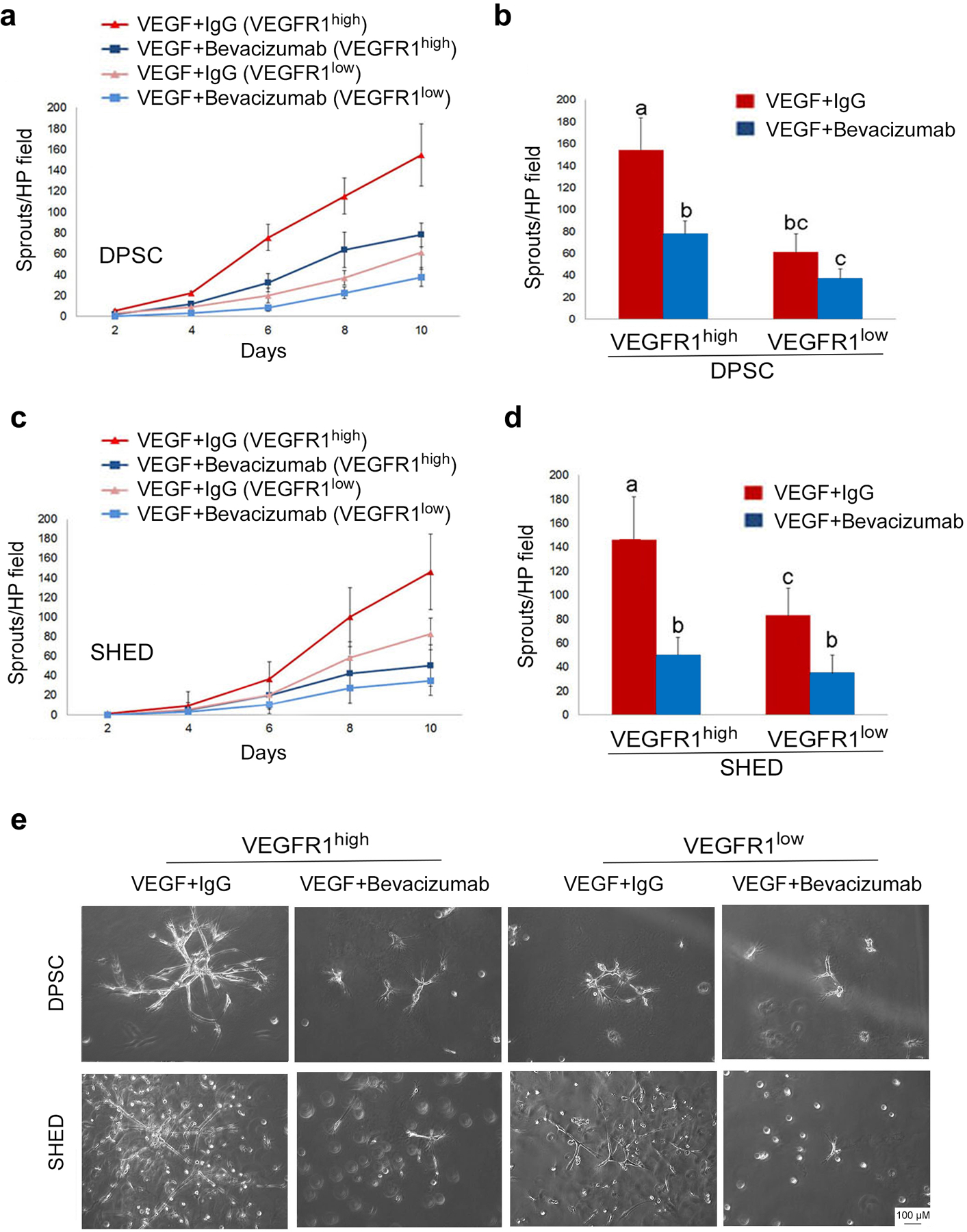
Dental pulp stem cells (DPSCs) constitute a unique group of cells endowed with multipotency, self-renewal, and capacity to regenerate the dental pulp tissue. While much has been learned about these cells in recent years, it is still unclear if each DPSC is multipotent or if unique sub-populations of DPSCs are "primed" to undergo specific differentiation paths. The purpose of the present study was to define whether a sub-population of DPSCs was uniquely primed to undergo vasculogenic differentiation. Permanent-tooth DPSCs or stem cells from human exfoliated deciduous teeth (SHED) were flow-sorted for vascular endothelial growth factor receptor 1 (VEGFR1) and exposed to vasculogenic differentiation medium, i.e., Microvascular-Endothelial-Cell-Growth-Medium-2-BulletKit™ supplemented with 50 ng/mL rhVEGF165 in the presence of 0 or 25 μg/mL anti-human VEGF antibody (bevacizumab; Genentech). In addition, sorted SHED (i.e., VEGFR1high or VEGFR1low) were seeded in biodegradable scaffolds and transplanted into the subcutaneous space of immunodeficient mice. Despite proliferating at a similar rate, VEGFR1high generated more in vitro sprouts than VEGFR1low cells (p < 0.05). Blockade of VEGF signaling with bevacizumab inhibited VEGFR1high-derived sprouts, demonstrating specificity of responses. Similarly, VEGFR1high SHED generated more blood vessels when transplanted into murine hosts than VEGFR1low cells (p < 0.05). Collectively, these data demonstrated that DPSCs contain a unique sub-population of cells defined by high VEGFR1 expression that are primed to differentiate into vascular endothelial cells. These data raise the possibility of purifying stem cells with high vasculogenic potential for regeneration of vascularized tissues or for vascular engineering in the treatment of ischemic conditions.
牙髓干细胞(DPSCs)构成了一组独特的细胞,具有多能性、自我更新和再生牙髓组织的能力。虽然近年来对这些细胞有了很多了解,但仍不清楚每个 DPSC 是否具有多能性,或者 DPSCs 的独特亚群是否“预先”具备进行特定分化途径的能力。本研究旨在确定 DPSCs 的亚群是否具有独特的血管生成分化潜能。对恒牙 DPSCs 或人脱落乳牙的干细胞(SHED)进行血管内皮生长因子受体 1(VEGFR1)的流式分选,并将其暴露于血管生成分化培养基中,即在微血管内皮细胞生长培养基-2 试剂盒(Microvascular-Endothelial-Cell-Growth-Medium-2-BulletKit™)中添加 50ng/ml rhVEGF165,并在存在 0 或 25μg/ml 抗人 VEGF 抗体(贝伐单抗;基因泰克)的情况下。此外,对分选的 SHED(即 VEGFR1high 或 VEGFR1low)进行种子处理,并移植到免疫缺陷小鼠的皮下空间。尽管 VEGFR1high 细胞的增殖速度相似,但它们在体外产生的芽比 VEGFR1low 细胞多(p<0.05)。用贝伐单抗阻断 VEGF 信号抑制了 VEGFR1high 衍生的芽,证明了反应的特异性。同样,当移植到小鼠宿主中时,VEGFR1high SHED 产生的血管比 VEGFR1low 细胞多(p<0.05)。综上所述,这些数据表明,DPSCs 包含一个独特的亚群,其特征是高 VEGFR1 表达,预先分化为血管内皮细胞。这些数据提出了一种可能性,即可以纯化具有高血管生成潜力的干细胞,用于血管化组织的再生或用于缺血性疾病治疗的血管工程。