Thompson Jeffrey R, Paganos Periklis, Benvenuto Giovanna, Arnone Maria Ina, Oliveri Paola
Department of Genetics, Evolution and Environment, University College London, Darwin Building, Gower Street, London, WC1E 6BT, UK.
UCL Center for Life's Origins and Evolution, London, UK.
Evodevo. 2021 Mar 16;12(1):3. doi: 10.1186/s13227-021-00174-1.
Understanding the molecular and cellular processes that underpin animal development are crucial for understanding the diversity of body plans found on the planet today. Because of their abundance in the fossil record, and tractability as a model system in the lab, skeletons provide an ideal experimental model to understand the origins of animal diversity. We herein use molecular and cellular markers to understand the growth and development of the juvenile sea urchin (echinoid) skeleton.
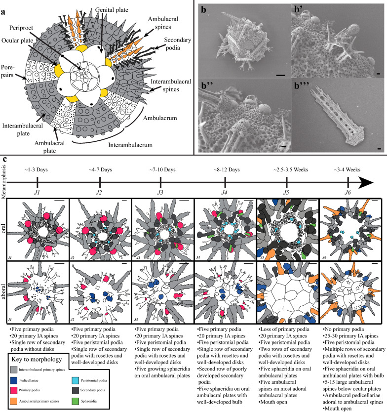
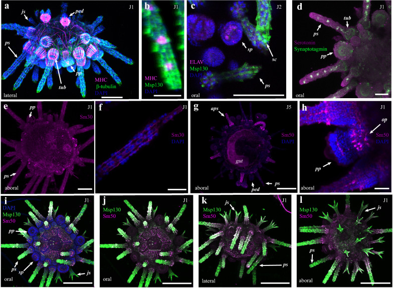
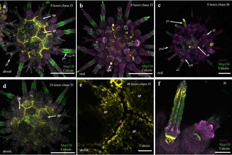
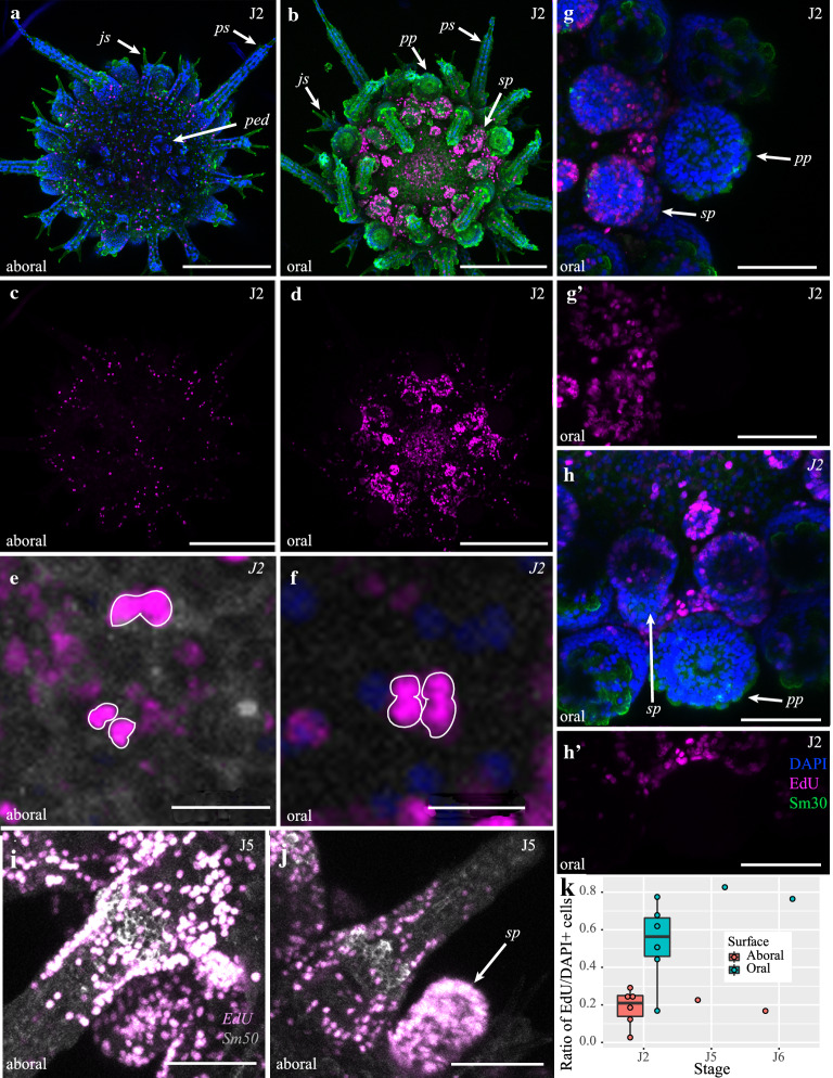
We developed a detailed staging scheme based off of the first ~ 4 weeks of post-metamorphic life of the regular echinoid Paracentrotus lividus. We paired this scheme with immunohistochemical staining for neuronal, muscular, and skeletal tissues, and fluorescent assays of skeletal growth and cell proliferation to understand the molecular and cellular mechanisms underlying skeletal growth and development of the sea urchin body plan.
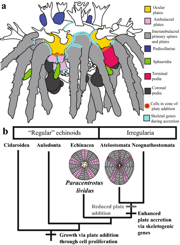
Our experiments highlight the role of skeletogenic proteins in accretionary skeletal growth and cell proliferation in the addition of new metameric tissues. Furthermore, this work provides a framework for understanding the developmental evolution of sea urchin body plans on macroevolutionary timescales.
了解支撑动物发育的分子和细胞过程对于理解当今地球上发现的身体结构多样性至关重要。由于骨骼在化石记录中数量丰富,且作为实验室中的模型系统易于处理,因此骨骼提供了一个理想的实验模型来理解动物多样性的起源。我们在此使用分子和细胞标记物来了解幼年海胆(海胆纲)骨骼的生长和发育。
我们基于规则海胆纲物种紫球海胆变态后约前4周的生活情况制定了详细的分期方案。我们将该方案与针对神经、肌肉和骨骼组织的免疫组织化学染色以及骨骼生长和细胞增殖的荧光测定相结合,以了解海胆身体结构骨骼生长和发育的分子和细胞机制。
我们的实验突出了成骨蛋白在新分节组织添加过程中增生性骨骼生长和细胞增殖中的作用。此外,这项工作为在宏观进化时间尺度上理解海胆身体结构的发育进化提供了一个框架。