Zhongshan Ophthalmic Center, State Key Laboratory of Ophthalmology, Sun Yat-Sen University, Guangzhou; Hainan Eye Hospital and Key Laboratory of Ophthalmology, Zhongshan Ophthalmic Center, Sun Yat-sen University, Haikou, People's Republic of China.
Zhongshan Ophthalmic Center, State Key Laboratory of Ophthalmology, Sun Yat-Sen University, Guangzhou, People's Republic of China.
Indian J Ophthalmol. 2021 Apr;69(4):865-870. doi: 10.4103/ijo.IJO_936_20.
Obtaining a better understanding of the pathogenesis of primary angle-closure disease (PACD) still requires studies that provide measurements of anterior and posterior biometric characteristics together and that assess the relationship between them.
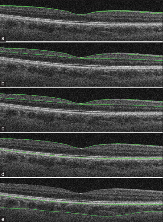
In total, 201 eyes were enrolled in this cross-sectional study: 50 normal controls, 49 primary angle-closure suspect (PACS), 38 primary angle closure (PAC), and 64 primary angle-closure glaucoma (PACG) eyes. The anterior and posterior structural features were measured by anterior segment optical coherence tomography and swept-source optical coherence tomography.
All PACD groups had smaller anterior chamber depth (ACD), anterior chamber area (ACA), anterior chamber volume (ACV), angle opening distance at 750 μm from the scleral spur (AOD750), trabecular-iris space area at 750 μm from the scleral spur (TISA750), and angle recess area (ARA), as well as a larger lens vault (LV), than controls (all P < 0.001). The PACS and PAC groups had thicker iris thickness at 750 μm from the scleral spur (IT750) than controls (P = 0.017 and P = 0.002, respectively). Choroidal thickness (CT) was not statistically different among normal, PACS, PAC, and PACG eyes. Univariate and multivariate linear regression analysis revealed a significant association between thinner IT750 and increased CT in PACD eyes (P = 0.031, univariate analysis; P = 0.008, multivariate analysis).
Thinner iris thickness was associated with increased CT in PACD eyes; however, the underlying mechanism needs further investigation.
为了更好地了解原发性闭角型青光眼(PAC)的发病机制,仍需要进行研究,这些研究需要同时提供前节和后节生物测量特征的测量值,并评估它们之间的关系。
本横断面研究共纳入 201 只眼:50 只正常对照眼、49 只原发性房角关闭可疑(PACS)眼、38 只原发性房角关闭(PAC)眼和 64 只原发性房角关闭性青光眼(PACG)眼。应用眼前节光学相干断层扫描和扫频源光学相干断层扫描测量前节和后节结构特征。
所有 PACD 组的前房深度(ACD)、前房面积(ACA)、前房容积(ACV)、巩膜突 750μm 处房角开放距离(AOD750)、巩膜突 750μm 处小梁虹膜空间面积(TISA750)和房角隐窝面积(ARA)均较小,晶状体拱高(LV)较大,与对照组相比差异均有统计学意义(均 P < 0.001)。PACS 和 PAC 组巩膜突 750μm 处虹膜厚度(IT750)比对照组厚(P = 0.017 和 P = 0.002)。正常、PACS、PAC 和 PACG 眼的脉络膜厚度(CT)无统计学差异。单变量和多变量线性回归分析显示,PAC 眼的 IT750 越薄,CT 越大(P = 0.031,单变量分析;P = 0.008,多变量分析)。
PAC 眼的虹膜厚度变薄与 CT 增加有关;但是,其潜在机制需要进一步研究。