Guo Huiying, Liu Wenjin, Li Haige, Yang Junwei
Department of Radiology, Second Affiliated Hospital of Nanjing Medical University, Nanjing, Jiangsu, People's Republic of China.
Center for Kidney Disease, Second Affiliated Hospital of Nanjing Medical University, Nanjing, Jiangsu, People's Republic of China.
Int J Nephrol Renovasc Dis. 2021 Mar 9;14:77-86. doi: 10.2147/IJNRD.S295025. eCollection 2021.
The current study aimed to depict intrinsic structural changes and the spontaneous brain activity patterns in voxel level in patients with end-stage renal disease (ESRD) undergoing hemodialysis (HD) by using diffusion-tensor imaging and resting-state functional magnetic resonance (MR) imaging with an amplitude of low-frequency fluctuations (ALFF) algorithm and their clinical relevance.
In the study, the diffusion-tensor imaging and resting-state functional MR imaging were performed in forty-two hemodialysis patients with ESRD and 42 healthy control subjects. Neuropsychological and laboratory tests were performed in all subjects. ALFF, fraction anisotropy (FA), and mean diffusivity (MD) values were compared between the two groups. Correlations between ALFF, FA or MD values, and clinical markers were analyzed.
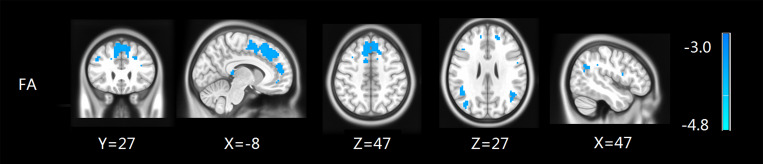
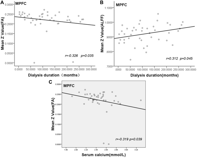
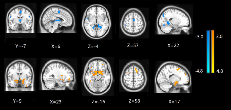
We found that ESRD patients exhibited significantly lower ALFF values in multiple areas, including medial frontal gyrus, limbic lobe, superior frontal gyrus, bilateral lingual gyri, occipital lobe, parahippocampal gyrus, precuneus, while increased ALFF values in medial frontal gyrus than healthy controls. FA values were decreased in medial frontal gyrus, parietal lobe, and left precuneus regions in the ESRD group compared with controls. Importantly, FA for the frontal and parietal lobes was negatively associated with the dialysis duration of ESRD patients, ALFF z-scores for the medial prefrontal cortex (MPFC) were positively correlated with the dialysis duration of ESRD patients and Serum calcium of ESRD patients negatively correlated with FA values in the frontal and parietal lobes (<0.05).
Our study revealed that both impaired brain structure and function in ESRD patients with routine hemodialysis distributed mainly in the parietal, temporal, and frontal lobes. ESRD patients have cognitive impairment and declined memory ability. Serum calcium and dialysis duration might be associated with the impairment of brain structure and function in patients with ESRD.
本研究旨在通过扩散张量成像和基于低频振幅波动(ALFF)算法的静息态功能磁共振成像,描绘终末期肾病(ESRD)接受血液透析(HD)患者体素水平的内在结构变化和自发脑活动模式及其临床相关性。
本研究对42例接受血液透析的ESRD患者和42名健康对照者进行了扩散张量成像和静息态功能磁共振成像。对所有受试者进行了神经心理学和实验室检查。比较了两组之间的ALFF、各向异性分数(FA)和平均扩散率(MD)值。分析了ALFF、FA或MD值与临床指标之间的相关性。
我们发现,ESRD患者在多个区域表现出显著较低的ALFF值,包括内侧前额叶回、边缘叶、额上回、双侧舌回、枕叶、海马旁回、楔前叶,而内侧前额叶回的ALFF值高于健康对照者。与对照组相比,ESRD组内侧前额叶回、顶叶和左侧楔前叶区域的FA值降低。重要的是,额叶和顶叶的FA与ESRD患者的透析时间呈负相关,内侧前额叶皮质(MPFC)的ALFF z评分与ESRD患者的透析时间呈正相关,ESRD患者的血清钙与额叶和顶叶的FA值呈负相关(<0.05)。
我们的研究表明,常规血液透析的ESRD患者脑结构和功能受损主要分布在顶叶、颞叶和额叶。ESRD患者存在认知障碍和记忆能力下降。血清钙和透析时间可能与ESRD患者脑结构和功能受损有关。