Su Huanhuan, Fu Shishun, Liu Mengchen, Yin Yi, Hua Kelei, Meng Shandong, Jiang Guihua, Quan Xianyue
Department of Radiology, Zhujiang Hospital, Southern Medical University, Guangzhou, China.
Department of Medical Imaging, Guangdong Second Provincial General Hospital, Guangzhou, China.
Front Neurol. 2022 Feb 9;12:801336. doi: 10.3389/fneur.2021.801336. eCollection 2021.
Using the amplitude of low-frequency fluctuation (ALFF) and functional connectivity (FC) algorithm to study the alteration of brain function in hemodialysis patients with end-stage renal disease (ESRD).
We recruited 20 patients with ESRD on regular hemodialysis and 17 healthy controls (HCs). All of the participants underwent resting-state fMRI (rs-fMRI), neuropsychological tests, and blood biochemical examination. The individual ALFF values between the two groups were tested by an independent sample -test. Then, we set the altered ALFF brain areas as seed regions of interest (ROIs), and FC analysis was used to investigate the functional integration patterns between the seed ROI and the voxels within the whole brain.
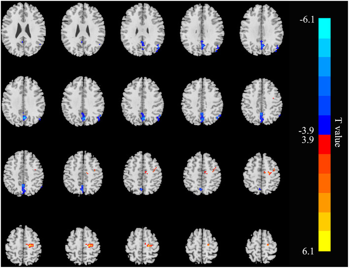
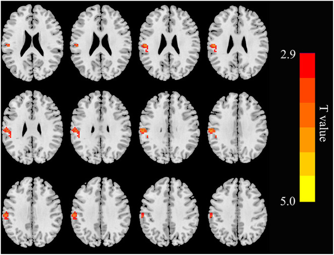
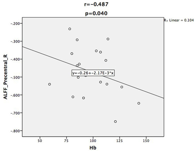
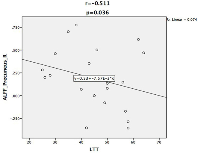
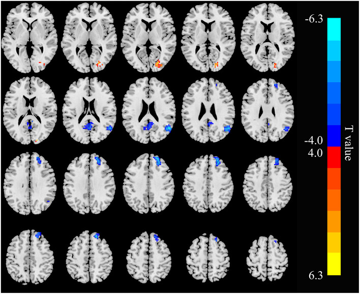
The ALFF values of the right precuneus and angular gyrus (RAG) in the ESRD group were lower than those in the HC subjects, but the right precentral gyrus showed higher ALFF values in patients. Hemoglobin (Hb) was negatively correlated with the ALFF values of the right precentral gyrus, and the ALFF values of the right precuneus were negatively correlated with line-tracing test (LTT) scores in patients with ESRD. Patients with ESRD show decreased connectivity between the RAG and the left precuneus, right superior frontal gyrus (RSFG), and the connectivity within the RAG was weak. In addition, FC in the RAG-right cuneus, right precuneus-left supramarginal gyrus was enhanced in the patient group.
Our research suggested that, in hemodialysis patients with ESRD, the brain areas with abnormal spontaneous brain activity and FC are mainly located in the default mode network (DMN) regions. Hb and the LTT results were correlated with abnormal spontaneous brain activity. These findings provide additional evidence to understand the possible underlying neuropathological mechanisms in patients with ESRD.
运用低频振幅(ALFF)和功能连接(FC)算法研究终末期肾病(ESRD)血液透析患者的脑功能改变。
我们招募了20例接受规律血液透析的ESRD患者和17名健康对照者(HCs)。所有参与者均接受静息态功能磁共振成像(rs-fMRI)、神经心理学测试及血液生化检查。两组间的个体ALFF值采用独立样本t检验。然后,将ALFF改变的脑区设为感兴趣种子区(ROIs),并运用FC分析研究种子ROI与全脑体素间的功能整合模式。
ESRD组右侧楔前叶和角回(RAG)的ALFF值低于HC组,但右侧中央前回在患者中显示出较高的ALFF值。血红蛋白(Hb)与右侧中央前回的ALFF值呈负相关,ESRD患者右侧楔前叶的ALFF值与线追踪试验(LTT)评分呈负相关。ESRD患者显示RAG与左侧楔前叶、右侧额上回(RSFG)之间的连接性降低,且RAG内的连接性较弱。此外,患者组中RAG-右侧楔叶、右侧楔前叶-左侧缘上回的FC增强。
我们的研究表明,在ESRD血液透析患者中,自发脑活动和FC异常的脑区主要位于默认模式网络(DMN)区域。Hb和LTT结果与异常的自发脑活动相关。这些发现为理解ESRD患者可能的潜在神经病理机制提供了更多证据。