West German Study Group, Ludwig-Weber-Strasse 15, 41061, Moenchengladbach, Germany.
Ev. Hospital Bethesda, Breast Center Niederrhein, Ludwig-Weber-Strasse 15, 41061, Moenchengladbach, Germany.
Breast Cancer Res. 2021 Mar 18;23(1):36. doi: 10.1186/s13058-021-01413-y.
Prediction of histological tumor size by post-neoadjuvant therapy (NAT) ultrasound and magnetic resonance imaging (MRI) was evaluated in different breast cancer subtypes.
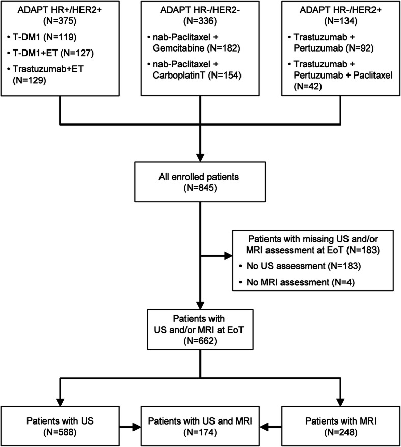
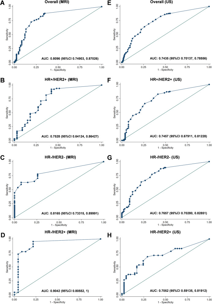
Imaging was performed after 12-week NAT in patients enrolled into three neoadjuvant WSG ADAPT subtrials. Imaging performance was analyzed for prediction of residual tumor measuring ≤10 mm and summarized using positive (PPV) and negative (NPV) predictive values.
A total of 248 and 588 patients had MRI and ultrasound, respectively. Tumor size was over- or underestimated by < 10 mm in 4.4% and 21.8% of patients by MRI and in 10.2% and 15.8% by ultrasound. Overall, NPV (proportion of correctly predicted tumor size ≤10 mm) of MRI and ultrasound was 0.92 and 0.83; PPV (correctly predicted tumor size > 10 mm) was 0.52 and 0.61. MRI demonstrated a higher NPV and lower PPV than ultrasound in hormone receptor (HR)-positive/human epidermal growth factor receptor 2 (HER2)-positive and in HR-/HER2+ tumors. Both methods had a comparable NPV and PPV in HR-/HER2- tumors.
In HR+/HER2+ and HR-/HER2+ breast cancer, MRI is less likely than ultrasound to underestimate while ultrasound is associated with a lower risk to overestimate tumor size. These findings may help to select the most optimal imaging approach for planning surgery after NAT.
Clinicaltrials.gov , NCT01815242 (registered on March 21, 2013), NCT01817452 (registered on March 25, 2013), and NCT01779206 (registered on January 30, 2013).
在不同乳腺癌亚型中,评估了新辅助治疗(NAT)后超声和磁共振成像(MRI)对组织学肿瘤大小的预测。
在三个新辅助 WSG ADAPT 子试验中招募的患者接受 12 周 NAT 后进行影像学检查。分析了影像学表现,以预测残留肿瘤大小≤10mm,并使用阳性(PPV)和阴性(NPV)预测值进行总结。
共有 248 例和 588 例患者分别进行了 MRI 和超声检查。MRI 和超声检查中,肿瘤大小低估<10mm的患者分别占 4.4%和 21.8%,高估<10mm的患者分别占 10.2%和 15.8%。总体而言,MRI 和超声的 NPV(预测肿瘤大小≤10mm的正确比例)分别为 0.92 和 0.83;PPV(正确预测肿瘤大小>10mm)分别为 0.52 和 0.61。在激素受体(HR)阳性/人表皮生长因子受体 2(HER2)阳性和 HR-/HER2+肿瘤中,MRI 的 NPV 高于超声,PPV 低于超声。在 HR-/HER2-肿瘤中,两种方法的 NPV 和 PPV 具有可比性。
在 HR+/HER2+和 HR-/HER2+乳腺癌中,MRI 不太可能低估肿瘤大小,而超声则不太可能高估肿瘤大小。这些发现可能有助于选择最适合新辅助治疗后手术计划的最佳影像学方法。
Clinicaltrials.gov ,NCT01815242(于 2013 年 3 月 21 日注册)、NCT01817452(于 2013 年 3 月 25 日注册)和 NCT01779206(于 2013 年 1 月 30 日注册)。