Xu Huimin, Zhao Yuanyuan, Suo Yuanzhen, Guo Yayu, Man Yi, Jing Yanping, He Xinqiang, Lin Jinxing
College of Biological Sciences, China Agricultural University, Beijing, 100193, China.
Beijing Advanced Innovation Center for Tree Breeding by Molecular Design, Beijing Forestry University, Beijing, 10083, China.
Plant Methods. 2021 Mar 19;17(1):29. doi: 10.1186/s13007-021-00730-9.
New cell wall imaging tools permit direct visualization of the molecular architecture of cell walls and provide detailed chemical information on wall polymers, which will aid efforts to use these polymers in multiple applications; however, detailed imaging and quantification of the native composition and architecture in the cell wall remains challenging.
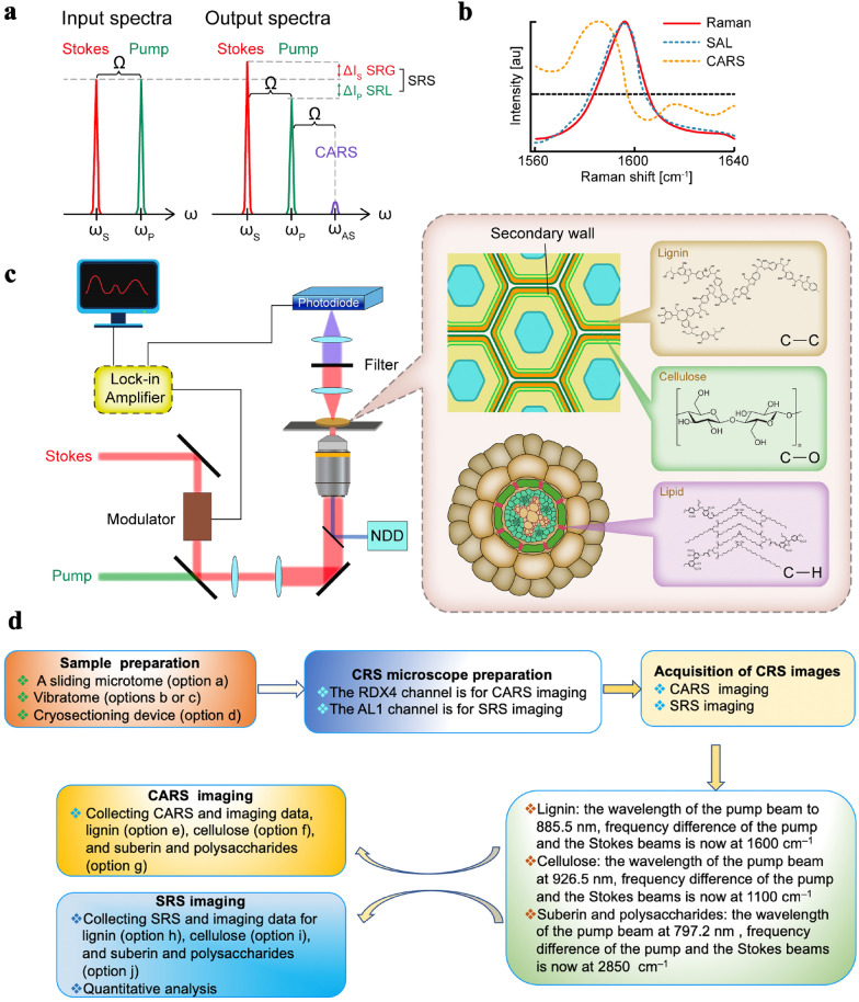
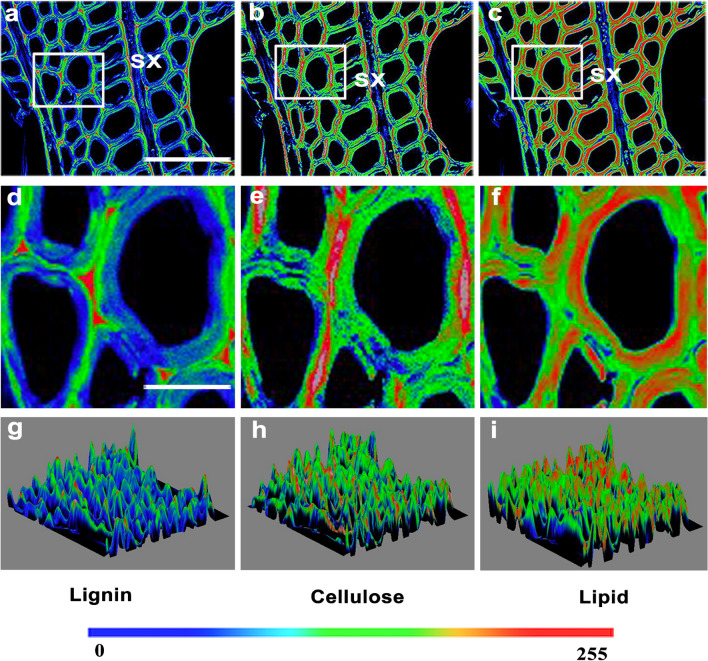
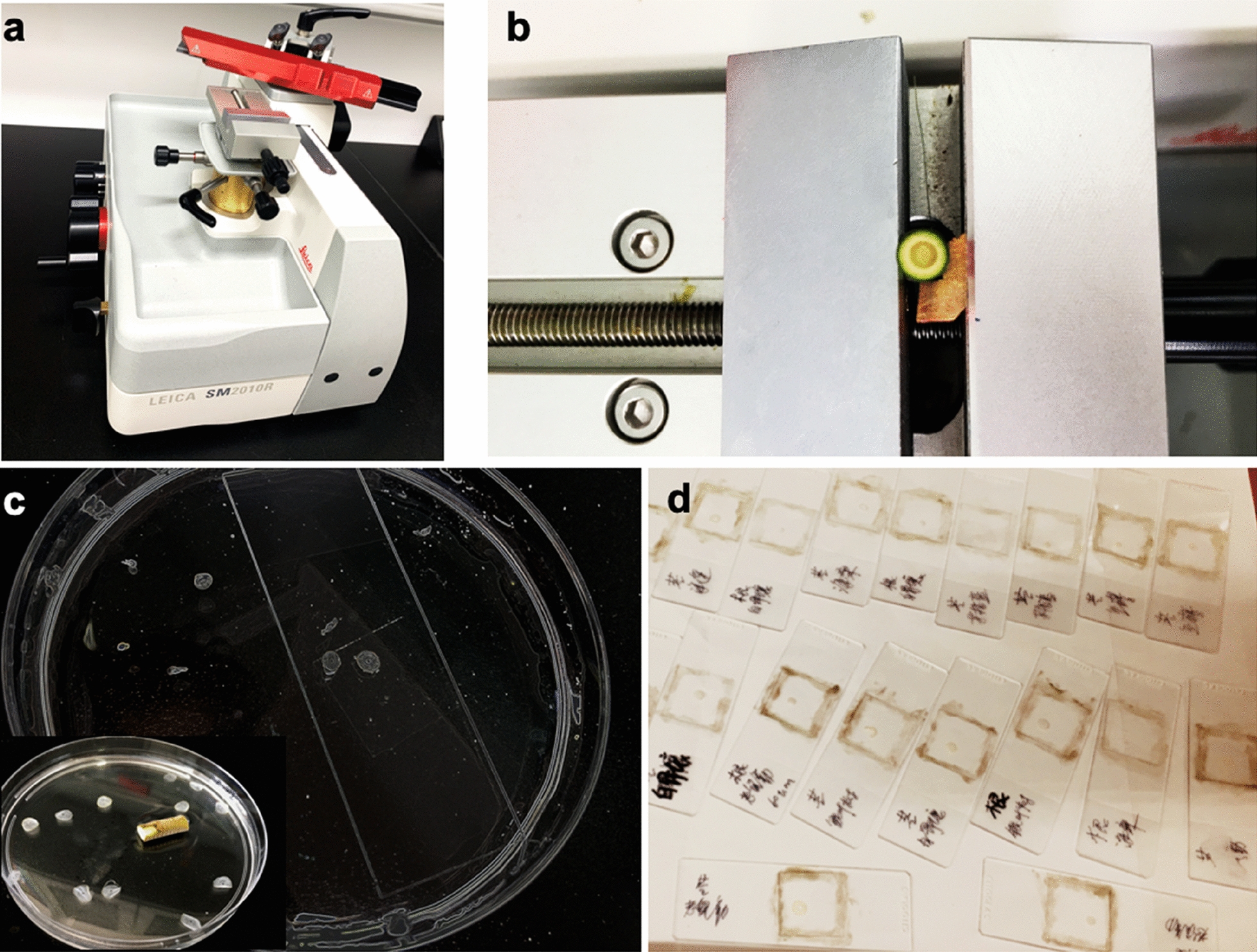
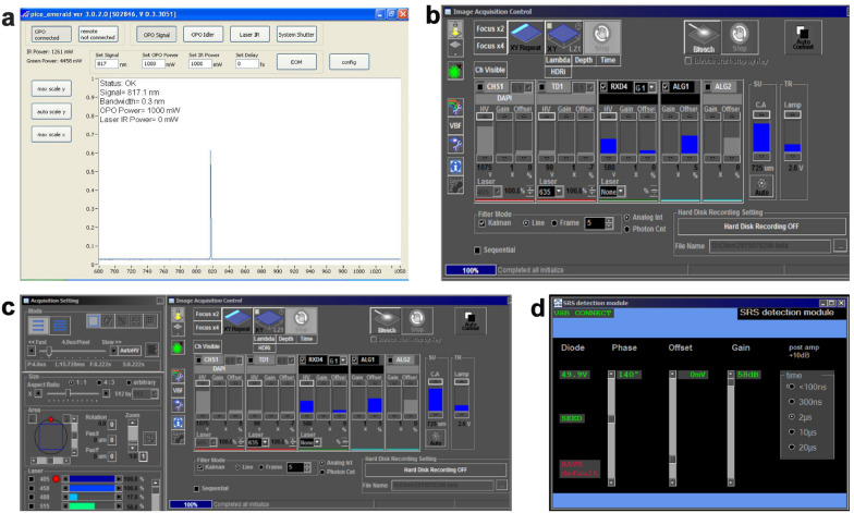
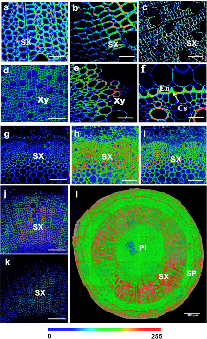
Here, we describe a label-free imaging technology, coherent Raman scattering (CRS) microscopy, including coherent anti-Stokes Raman scattering (CARS) microscopy and stimulated Raman scattering (SRS) microscopy, which can be used to visualize the major structures and chemical composition of plant cell walls. We outline the major steps of the procedure, including sample preparation, setting the mapping parameters, analysis of spectral data, and image generation. Applying this rapid approach will help researchers understand the highly heterogeneous structures and organization of plant cell walls.
This method can potentially be incorporated into label-free microanalyses of plant cell wall chemical composition based on the in situ vibrations of molecules.
新型细胞壁成像工具能够直接观察细胞壁的分子结构,并提供有关细胞壁聚合物的详细化学信息,这将有助于在多种应用中使用这些聚合物;然而,对细胞壁中天然成分和结构进行详细成像和定量分析仍然具有挑战性。
在此,我们描述了一种无标记成像技术——相干拉曼散射(CRS)显微镜,包括相干反斯托克斯拉曼散射(CARS)显微镜和受激拉曼散射(SRS)显微镜,可用于观察植物细胞壁的主要结构和化学成分。我们概述了该过程的主要步骤,包括样品制备、设置映射参数、光谱数据分析和图像生成。应用这种快速方法将有助于研究人员了解植物细胞壁高度异质的结构和组织。
基于分子的原位振动,该方法有可能被纳入植物细胞壁化学成分的无标记微分析中。