• 文献检索
  • 文档翻译
  • 深度研究
  • 学术资讯
  • Suppr Zotero 插件Zotero 插件
  • 邀请有礼
  • 套餐&价格
  • 历史记录
应用&插件
Suppr Zotero 插件Zotero 插件浏览器插件Mac 客户端Windows 客户端微信小程序
定价
高级版会员购买积分包购买API积分包
服务
文献检索文档翻译深度研究API 文档MCP 服务
关于我们
关于 Suppr公司介绍联系我们用户协议隐私条款
关注我们

Suppr 超能文献

核心技术专利:CN118964589B侵权必究
粤ICP备2023148730 号-1Suppr @ 2026

文献检索

告别复杂PubMed语法,用中文像聊天一样搜索,搜遍4000万医学文献。AI智能推荐,让科研检索更轻松。

立即免费搜索

文件翻译

保留排版,准确专业,支持PDF/Word/PPT等文件格式,支持 12+语言互译。

免费翻译文档

深度研究

AI帮你快速写综述,25分钟生成高质量综述,智能提取关键信息,辅助科研写作。

立即免费体验

云南西双版纳 8 例以肺部肿块为主要表现的成人并殖吸虫病误诊分析。

Analysis of the misdiagnosis of 8 adult cases of paragonimiasis with lung masses as the main manifestation in Xishuangbanna, Yunnan.

机构信息

The Second Affiliated Hospital of Kunming Medical University, No. 374, Dianmian Road, Kunming, 651010, Yunnan, China.

Oncology Department, People's hospital of Xishuangbanna Dai Autonomous Prefecture, Xishuangbanna, Jinghong, Yunnan, China.

出版信息

J Cardiothorac Surg. 2021 Mar 19;16(1):28. doi: 10.1186/s13019-021-01408-y.

DOI:10.1186/s13019-021-01408-y
PMID:33741016
原文链接:https://pmc.ncbi.nlm.nih.gov/articles/PMC7977500/
Abstract

OBJECTIVE

To summarize the clinical characteristics of adult cases of paragonimiasis with lung masses as the main manifestation in Xishuangbanna, Yunnan Province, analyze the causes of misdiagnosis, and improve the levels of clinical diagnosis and treatment.

METHOD

We conducted a retrospective analysis of the clinical data and diagnosis and treatment of 8 adult cases of paragonimiasis with lung masses as the main manifestation that were diagnosed in the Oncology Department of People's hospital of Xishuangbanna Dai Autonomous Prefecture from July 2014 to July 2019.

RESULT

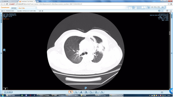
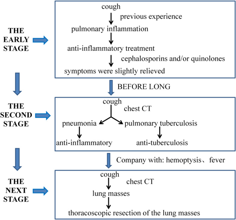
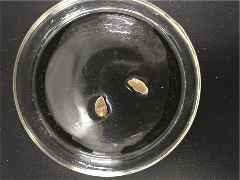
All 8 patients were from epidemic paragonimiasis areas and had a confirmed history of consuming uncooked freshwater crabs. The clinical manifestations were mainly fever, dry cough, and chest pain. The disease durations were long, and peripheral blood eosinophil counts were elevated. The cases had been misdiagnosed as pneumonia or pulmonary tuberculosis. After years of anti-inflammatory or anti-tuberculosis treatment, the symptoms had not improved significantly. Patients eventually sought treatment from the oncology department for hemoptysis. Chest computed tomography showed patchy consolidation in the lungs, with nodules, lung masses, and enlarged mediastinal lymph nodes.

CONCLUSION

Paragonimiasis is a food-borne parasitic disease. Early clinical manifestations and auxiliary examination results are nonspecific. The parasite most often invades the lungs, and the resulting disease is often misdiagnosed as pneumonia, pulmonary tuberculosis, or lung cancer (Acta Trop 199: 05074, 2019). To avoid misdiagnosis, clinicians should inquire, in detail, about residence history and history of unclean food and exposure to infected water and make an early diagnosis based on the inquired information and imaging examination results. For patients who have been diagnosed with pneumonia or pulmonary tuberculosis and whose symptoms do not improve significantly after anti-inflammatory or anti-tuberculosis treatments, their epidemiological history should be traced to further conduct differential diagnosis and avoid misdiagnosis.

摘要

目的

总结云南省西双版纳州以肺部肿块为主要表现的成人并殖吸虫病的临床特点,分析误诊原因,提高临床诊治水平。

方法

回顾性分析 2014 年 7 月至 2019 年 7 月在西双版纳州人民医院肿瘤科诊断的 8 例以肺部肿块为主要表现的成人并殖吸虫病的临床资料及诊治情况。

结果

8 例患者均来自并殖吸虫病流行区,有明确生食淡水蟹史,临床表现主要为发热、干咳、胸痛,病程长,外周血嗜酸性粒细胞计数升高,均误诊为肺炎或肺结核,经多年抗炎或抗结核治疗,症状无明显改善,因咯血就诊于肿瘤科,胸部 CT 表现为肺部斑片状实变影,伴有结节、肿块及纵隔淋巴结肿大。

结论

并殖吸虫病是一种食源性寄生虫病,早期临床表现和辅助检查结果不具特异性,寄生虫多侵犯肺部,导致的疾病常被误诊为肺炎、肺结核或肺癌(Acta Trop 199:05074,2019)。为避免误诊,临床医生应详细询问居住史、不洁食物史和接触受感染水的情况,并根据询问信息和影像学检查结果做出早期诊断。对于诊断为肺炎或肺结核且抗炎或抗结核治疗后症状无明显改善的患者,应追查其流行病学史,进一步进行鉴别诊断,避免误诊。

https://cdn.ncbi.nlm.nih.gov/pmc/blobs/08a3/7980663/9a12bba0690a/13019_2021_1408_Fig6_HTML.jpg
https://cdn.ncbi.nlm.nih.gov/pmc/blobs/08a3/7980663/1b5187ff7eed/13019_2021_1408_Fig1_HTML.jpg
https://cdn.ncbi.nlm.nih.gov/pmc/blobs/08a3/7980663/86afc3c1af4b/13019_2021_1408_Fig2_HTML.jpg
https://cdn.ncbi.nlm.nih.gov/pmc/blobs/08a3/7980663/880db27795b1/13019_2021_1408_Fig3_HTML.jpg
https://cdn.ncbi.nlm.nih.gov/pmc/blobs/08a3/7980663/45218c131db4/13019_2021_1408_Fig4_HTML.jpg
https://cdn.ncbi.nlm.nih.gov/pmc/blobs/08a3/7980663/1754c328b262/13019_2021_1408_Fig5_HTML.jpg
https://cdn.ncbi.nlm.nih.gov/pmc/blobs/08a3/7980663/9a12bba0690a/13019_2021_1408_Fig6_HTML.jpg

相似文献

1
Analysis of the misdiagnosis of 8 adult cases of paragonimiasis with lung masses as the main manifestation in Xishuangbanna, Yunnan.云南西双版纳 8 例以肺部肿块为主要表现的成人并殖吸虫病误诊分析。
J Cardiothorac Surg. 2021 Mar 19;16(1):28. doi: 10.1186/s13019-021-01408-y.
2
A retrospective clinical analysis of pediatric paragonimiasis in a Chinese children's hospital from 2011 to 2019.2011 年至 2019 年中国某儿童医院儿童并殖吸虫病的回顾性临床分析。
Sci Rep. 2021 Jan 21;11(1):2005. doi: 10.1038/s41598-021-81694-7.
3
Pleural fluid characteristics of pleuropulmonary paragonimiasis masquerading as pleural tuberculosis.伪装成胸膜结核的肺吸虫病的胸腔积液特征
Korean J Intern Med. 2015 Jan;30(1):56-61. doi: 10.3904/kjim.2015.30.1.56. Epub 2014 Dec 30.
4
Cerebral paragonimiasis: a retrospective analysis of 27 cases.脑型肺吸虫病:27例回顾性分析
J Neurosurg Pediatr. 2015 Jan;15(1):101-6. doi: 10.3171/2014.10.PEDS14208.
5
A rare case of paragonimiasis miyazakii with lung involvement diagnosed 7 years after infection: A case report and literature review.1例宫崎并殖吸虫病累及肺部且感染7年后才确诊的罕见病例:病例报告及文献复习
Parasitol Int. 2015 Oct;64(5):274-80. doi: 10.1016/j.parint.2015.02.009. Epub 2015 Mar 11.
6
A pulmonary paragonimiasis case mimicking metastatic pulmonary tumor.一例酷似转移性肺肿瘤的肺吸虫病病例。
Korean J Parasitol. 2011 Mar;49(1):69-72. doi: 10.3347/kjp.2011.49.1.69. Epub 2011 Mar 18.
7
[Paragonimiasis of the lung].[肺吸虫病]
Z Erkr Atmungsorgane. 1987;168(3):265-72.
8
A fluke occurrence of paramount significance.一个具有至关重要意义的侥幸事件。
J Postgrad Med. 2011 Jul-Sep;57(3):255-6. doi: 10.4103/0022-3859.85225.
9
Coinfection of Pulmonary Paragonimiasis and Pulmonary Tuberculosis in Thailand.泰国肺吸虫病与肺结核的合并感染
J Med Assoc Thai. 2016 Nov;99 Suppl 8:S231-S236.
10
[Clinicopathologic analysis of paragonimiasis].
Zhonghua Bing Li Xue Za Zhi. 2004 Apr;33(2):117-9.

引用本文的文献

1
Pericardial effusion in pediatric paragonimiasis: Surgery may not be necessary in cases of moderate to large effusion-a retrospective study.小儿肺吸虫病的心包积液:中至大量积液病例可能无需手术——一项回顾性研究
PLoS Negl Trop Dis. 2025 Apr 24;19(4):e0013023. doi: 10.1371/journal.pntd.0013023. eCollection 2025 Apr.
2
Analysis of clinical characteristics of 213 clinically diagnosed pediatric patients with paragonimiasis from Yunnan, China.对来自中国云南的213例临床诊断为肺吸虫病的儿科患者的临床特征进行分析。
BMC Infect Dis. 2025 Mar 24;25(1):397. doi: 10.1186/s12879-025-10776-y.
3
Paragonimiasis.

本文引用的文献

1
Development of point-of-care testing tool using immunochromatography for rapid diagnosis of human paragonimiasis.利用免疫层析法开发即时检测工具,用于快速诊断人体肺吸虫病。
Acta Trop. 2020 Mar;203:105325. doi: 10.1016/j.actatropica.2019.105325. Epub 2019 Dec 28.
2
Multiple cavitary lung lesions on CT: imaging findings to differentiate between malignant and benign etiologies.CT 上多发空洞性肺病变:鉴别良恶性病因的影像学特征。
J Bras Pneumol. 2019 Dec 20;46(2):e20190024. doi: 10.36416/1806-3756/e20190024. eCollection 2020.
3
Mini-FLOTAC as an alternative, non-invasive diagnostic tool for Schistosoma mansoni and other trematode infections in wildlife reservoirs.
并殖吸虫病。
Adv Exp Med Biol. 2024;1454:203-238. doi: 10.1007/978-3-031-60121-7_6.
4
What Findings on Chest CTs Can Delay Diagnosis of Pleuropulmonary Paragonimiasis?胸部 CT 有哪些发现可能会延误肺吸虫病的诊断?
Tomography. 2022 Jun 9;8(3):1493-1502. doi: 10.3390/tomography8030122.
5
Lung flukes of the genus : ancient and re-emerging pathogens.肺吸虫属的吸虫:古老且再现的病原体。
Parasitology. 2022 Sep;149(10):1286-1295. doi: 10.1017/S0031182022000300. Epub 2022 Mar 16.
6
Morphological and Molecular Characterization of Species Isolated from Freshwater Crabs in Southern Yunnan, China.中国云南南部淡水蟹分离物种的形态学和分子特征
J Trop Med. 2021 Dec 31;2021:5646291. doi: 10.1155/2021/5646291. eCollection 2021.
微型-FLOTAC 作为一种替代的、非侵入性的诊断工具,用于检测野生动物宿主中的曼氏血吸虫和其他吸虫感染。
Parasit Vectors. 2019 Sep 16;12(1):439. doi: 10.1186/s13071-019-3613-6.
4
Paragonimiasis.并殖吸虫病。
Adv Exp Med Biol. 2019;1154:105-138. doi: 10.1007/978-3-030-18616-6_5.
5
Paragonimus and paragonimiasis in Asia: An update.亚洲的并殖吸虫和并殖吸虫病:最新进展。
Acta Trop. 2019 Nov;199:105074. doi: 10.1016/j.actatropica.2019.105074. Epub 2019 Jul 8.
6
[Lung abscess: Diagnosis and management].[肺脓肿:诊断与治疗]
Rev Mal Respir. 2019 Jun;36(6):707-719. doi: 10.1016/j.rmr.2018.07.010. Epub 2019 Jun 13.
7
Progress on the development of rapid diagnostic tests for foodborne neglected zoonotic helminthiases: A systematic review.食源性被忽视的人畜共患蠕虫病快速诊断检测方法的研发进展:一项系统综述
Acta Trop. 2019 Jun;194:135-147. doi: 10.1016/j.actatropica.2019.03.030. Epub 2019 Apr 1.
8
Estimation of disability weight for paragonimiasis: a systematic analysis.估算并分析并殖吸虫病的失能权重:一项系统分析。
Infect Dis Poverty. 2018 Oct 19;7(1):110. doi: 10.1186/s40249-018-0485-5.
9
Paragonimus and paragonimiasis in West and Central Africa: unresolved questions.西非和中非的并殖吸虫及并殖吸虫病:未解决的问题
Parasitology. 2018 Nov;145(13):1748-1757. doi: 10.1017/S0031182018001439. Epub 2018 Sep 13.
10
[Pulmonary larval paragonimiasis mimicking lung cancer].[酷似肺癌的肺幼虫型肺吸虫病]
Arkh Patol. 2018;80(2):60-63. doi: 10.17116/patol201880260-63.