Department of Radiation Oncology, University Hospital Muenster, Albert-Schweitzer-Campus 1, 48149, Muenster, Germany.
West German Cancer Center, Muenster and Essen, Germany.
Ann Nucl Med. 2021 May;35(5):628-638. doi: 10.1007/s12149-021-01606-7. Epub 2021 Mar 19.
The objective of this study was to assess the accuracy of Ga-PSMA-11 PET/MRI, F-PSMA-1007 PET/CT, Ga-PSMA-11 PET/CT, and multiparametric (mp)MRI for the delineating of dominant intraprostatic lesions (IPL).
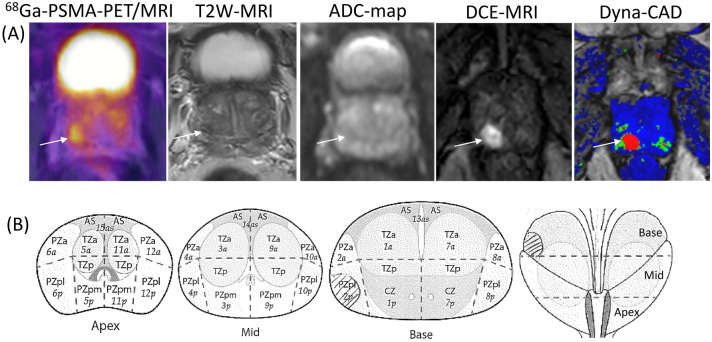
35 patients with organ-confined prostate cancer who were assigned to definitive radiotherapy (RT) were divided into three groups based on imaging techniques: Ga-PSMA-PET/MRI (n = 9), F-PSMA-PET/CT (n = 16) and Ga-PSMA-PET/CT (n = 10). All patients without PSMA-PET/MRI received an additional mpMRI. PSMA-PET-based automatic isocontours and manual contours of the dominant IPLs were generated for each modality. The biopsy results were then used to validate whether any of the prostate biopsies were positive in the marked lesion using Dice similarity coefficient (DSC), Youden index (YI), sensitivity and specificity. Factors that can predict the accuracy of IPLs contouring were analysed.
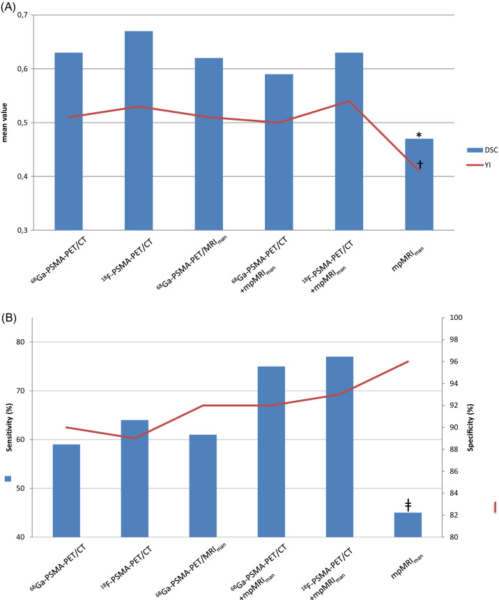
Diagnostic performance was significantly superior both for manual and automatic IPLs contouring using Ga-PSMA-PET/MRI (DSC/YI SUV-0.62/0.51), F-PSMA-PET/CT (DSC/YI SUV-0.67/0.53) or Ga-PSMA-PET/CT (DSC/YI SUV-0.63/0.51) compared to mpMRI (DSC/YI-0.47/0.41; p < 0.001). The accuracy for delineating IPLs was not improved by combination of PET/CT and mpMRI images compared to PET/CT alone. Significantly superior diagnostic accuracy was found for large prostate lesions (at least 15% from the prostate volume) and higher Gleason score (at least 7b) comparing to smaller lesions with lower GS.
IPL localization was significantly improved when using PSMA-imaging procedures compared to mpMRI. No significant difference for delineating IPLs was found between hybrid method PSMA-PET/MRI and PSMA-PET/CT. PSMA-based imaging technique should be considered for the diagnostics of IPLs and focal treatment modality.
本研究旨在评估 Ga-PSMA-11 PET/MRI、F-PSMA-1007 PET/CT、Ga-PSMA-11 PET/CT 和多参数(mp)MRI 对勾画主导性前列腺内病变(IPL)的准确性。
35 例局限于前列腺癌患者,根据影像学技术分为三组:Ga-PSMA-PET/MRI(n=9)、F-PSMA-PET/CT(n=16)和 Ga-PSMA-PET/CT(n=10)。所有未行 PSMA-PET/MRI 检查的患者均加做 mpMRI。对每种方法的 Ga-PSMA-PET 自动勾画和主导 IPL 手动勾画进行勾画。然后,使用 Dice 相似系数(DSC)、Youden 指数(YI)、敏感度和特异度来验证活检结果是否在标记病变中有阳性前列腺活检。分析可预测 IPL 勾画准确性的因素。
与 mpMRI 相比,Ga-PSMA-PET/MRI(DSC/YI SUV-0.62/0.51)、F-PSMA-PET/CT(DSC/YI SUV-0.67/0.53)或 Ga-PSMA-PET/CT(DSC/YI SUV-0.63/0.51)的手动和自动勾画对主导 IPL 勾画的诊断性能明显更高(DSC/YI SUV-0.63/0.51;p<0.001)。与单独使用 PET/CT 相比,PET/CT 联合 mpMRI 并不能提高 IPL 勾画的准确性。与较小、GS 较低的病变相比,较大的前列腺病变(至少占前列腺体积的 15%)和较高的 Gleason 评分(至少 7b)的诊断准确性明显更高。
与 mpMRI 相比,PSMA 成像程序可显著提高 IPL 定位。在勾画 IPL 方面,混合方法 PSMA-PET/MRI 和 PSMA-PET/CT 之间没有发现显著差异。PSMA 成像技术应考虑用于 IPL 的诊断和局灶治疗方式。