Christian Doppler Laboratory for Medical Radiation Research for Radiation Oncology, Medical University of Vienna, Vienna, Austria.
Department of Radiotherapy, Comprehensive Cancer Center, Medical University of Vienna/Vienna General Hospital, Vienna, Austria.
Strahlenther Onkol. 2019 May;195(5):402-411. doi: 10.1007/s00066-018-1402-3. Epub 2018 Nov 26.
Accurate prostate cancer (PCa) detection is essential for planning focal external beam radiotherapy (EBRT). While biparametric MRI (bpMRI) including T2-weighted (T2w) and diffusion-weighted images (DWI) is an accurate tool to localize PCa, its value is less clear in the case of additional androgen deprivation therapy (ADT). The aim of this study was to investigate the value of a textural feature (TF) approach on bpMRI analysis in prostate cancer patients with and without neoadjuvant ADT with respect to future dose-painting applications.
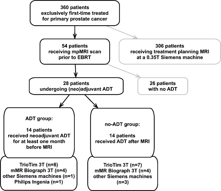
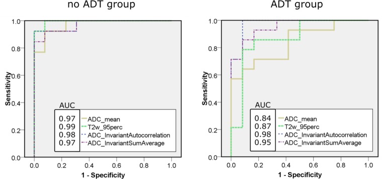
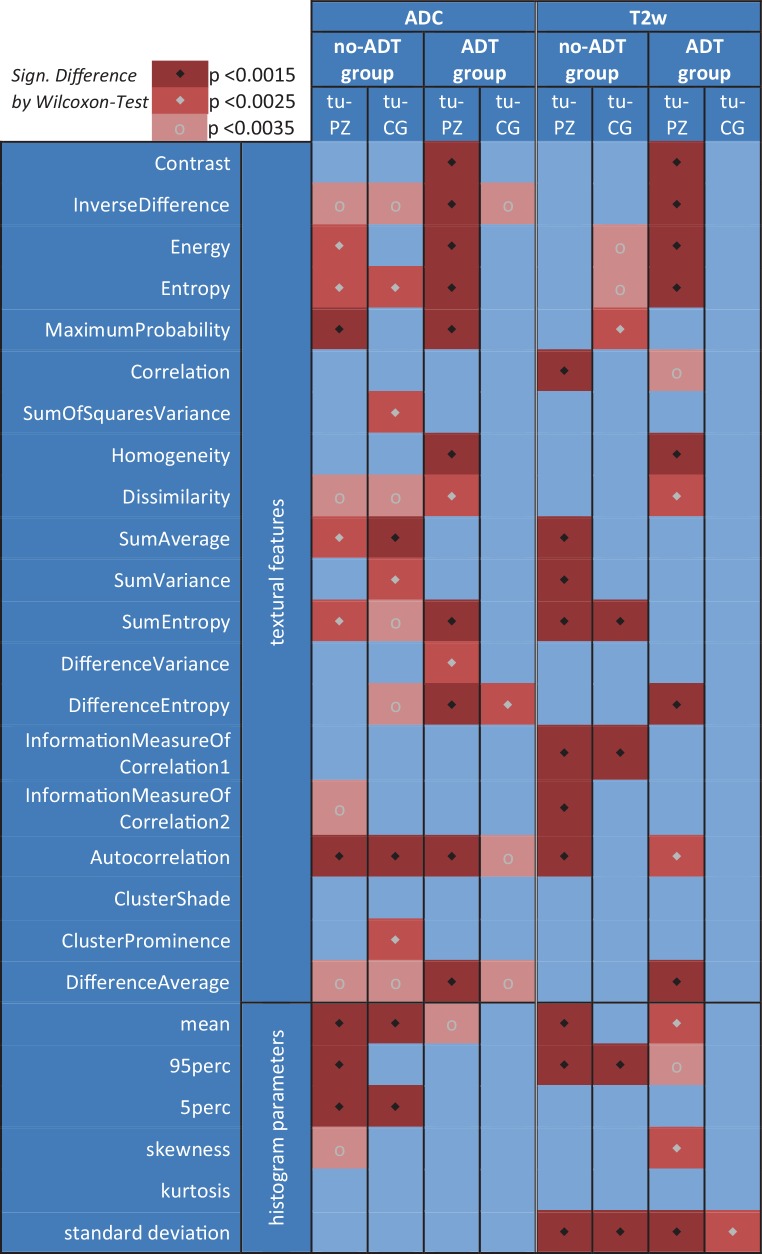
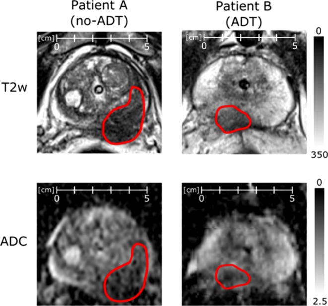
28 PCa patients (54-80 years) with (n = 14) and without (n = 14) ADT who underwent bpMRI with T2w and DWI were analyzed retrospectively. Lesions, central gland (CG), and peripheral zone (PZ) were delineated by an experienced urogenital radiologist based on localized pre-therapeutic histopathology. Histogram parameters and 20 Haralick TF were calculated. Regional differences (i. e., tumor vs. PZ, tumor vs. CG) were analyzed for all imaging parameters. Receiver-operating characteristic (ROC) analysis was performed to measure diagnostic performance to distinguish PCa from benign prostate tissue and to identify the features with best discriminative power in both patient groups.
The obtained sensitivities were equivalent or superior when utilizing the TF in the no-ADT group, while specificity was higher for the histogram parameters. However, in the ADT group, TF outperformed the conventional histogram parameters in both specificity and sensitivity. Rule-in and rule-out criteria for ADT patients could exclusively be defined with the aid of TF.
The TF approach has the potential for quantitative image-assisted boost volume delineation in PCa patients even if they are undergoing neoadjuvant ADT.
准确检测前列腺癌(PCa)对于规划局灶性外束放射治疗(EBRT)至关重要。虽然包括 T2 加权(T2w)和弥散加权成像(DWI)的双参数 MRI(bpMRI)是定位 PCa 的准确工具,但在附加雄激素剥夺治疗(ADT)的情况下,其价值不太明确。本研究旨在探讨在接受和不接受新辅助 ADT 的 PCa 患者的 bpMRI 分析中,纹理特征(TF)方法在未来剂量描绘应用中的价值。
回顾性分析了 28 例接受 bpMRI(包括 T2w 和 DWI)的 PCa 患者(54-80 岁),其中 14 例患者接受了 ADT,14 例患者未接受 ADT。一名有经验的泌尿生殖放射科医生根据局部治疗前的组织病理学对病变、中央腺体(CG)和外周带(PZ)进行了勾画。计算了直方图参数和 20 个 Haralick TF。对所有成像参数进行了区域差异(即肿瘤与 PZ、肿瘤与 CG)分析。进行了接收者操作特征(ROC)分析,以衡量区分 PCa 与良性前列腺组织的诊断性能,并确定在两组患者中具有最佳鉴别力的特征。
在无 ADT 组中使用 TF 时,获得的敏感性相等或更高,而在 ADT 组中,特异性更高。然而,在 ADT 组中,TF 在特异性和敏感性方面均优于传统的直方图参数。仅借助 TF 即可为 ADT 患者定义纳入和排除标准。
即使患者正在接受新辅助 ADT,TF 方法也有可能用于 PCa 患者的定量图像辅助增敏体积勾画。