Department of Radiology, Miller School of Medicine University of Miami, Miami, FL, USA.
Research and Development, MIM software Inc., Cleveland, OH, USA.
Strahlenther Onkol. 2019 Feb;195(2):121-130. doi: 10.1007/s00066-018-1348-5. Epub 2018 Aug 23.
The aim of this study was to evaluate an automatic multi-atlas-based segmentation method for generating prostate, peripheral (PZ), and transition zone (TZ) contours on MRIs with and without fat saturation (±FS), and compare MRIs from different vendor MRI systems.
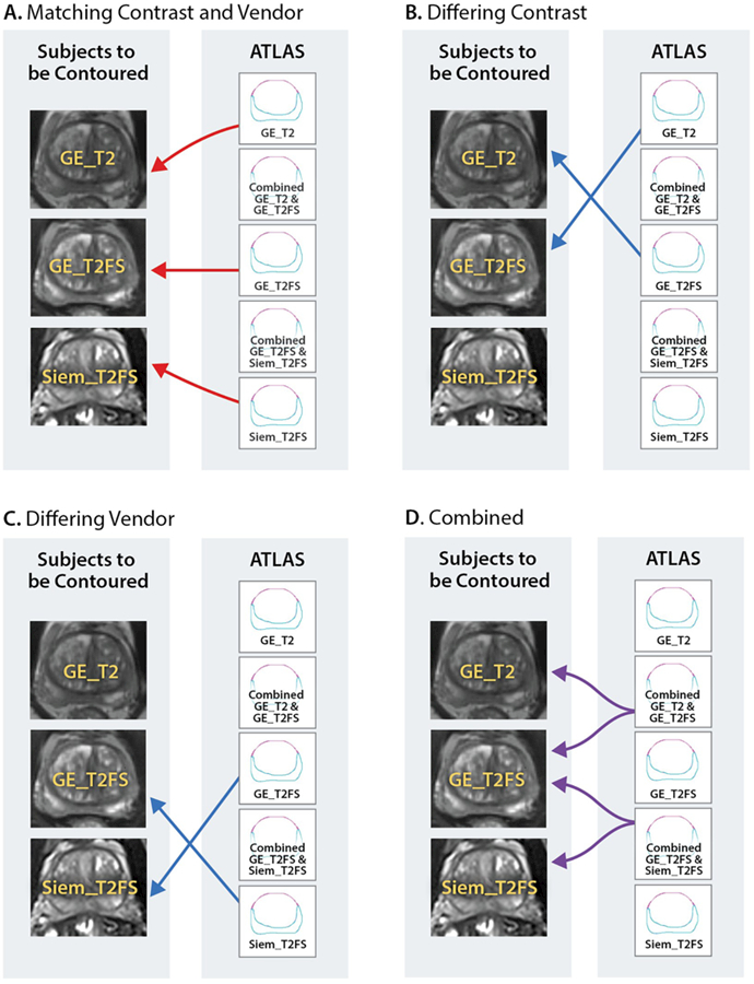
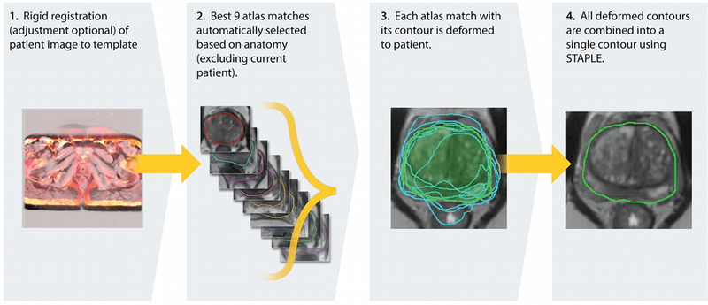
T2-weighted (T2) and fat-saturated (T2FS) MRIs were acquired on 3T GE (GE, Waukesha, WI, USA) and Siemens (Erlangen, Germany) systems. Manual prostate and PZ contours were used to create atlas libraries. As a test MRI is entered, the procedure for atlas segmentation automatically identifies the atlas subjects that best match the test subject, followed by a normalized intensity-based free-form deformable registration. The contours are transformed to the test subject, and Dice similarity coefficients (DSC) and Hausdorff distances between atlas-generated and manual contours were used to assess performance.
Three atlases were generated based on GE_T2 (n = 30), GE_T2FS (n = 30), and Siem_T2FS (n = 31). When test images matched the contrast and vendor of the atlas, DSCs of 0.81 and 0.83 for T2 ± FS were obtained (baseline performance). Atlases performed with higher accuracy when segmenting (i) T2FS vs. T2 images, likely due to a superior contrast between prostate vs. surrounding tissue; (ii) prostate vs. zonal anatomy; (iii) in the mid-gland vs. base and apex. Atlases performance declined when tested with images with differing contrast and MRI vendor. Conversely, combined atlases showed similar performance to baseline.
The MRI atlas-based segmentation method achieved good results for prostate, PZ, and TZ compared to expert contoured volumes. Combined atlases performed similarly to matching atlas and scan type. The technique is fast, fully automatic, and implemented on commercially available clinical platform.
本研究旨在评估一种基于自动多图谱的分割方法,用于生成有和无脂肪饱和(±FS)的 MRI 上的前列腺、外周区(PZ)和移行区(TZ)轮廓,并比较来自不同供应商 MRI 系统的 MRI。
在 3T 的 GE(美国威斯康星州沃基肖的 GE)和西门子(德国埃尔朗根)系统上采集 T2 加权(T2)和脂肪饱和(T2FS)MRI。手动前列腺和 PZ 轮廓用于创建图谱库。当输入测试 MRI 时,图谱分割过程会自动识别与测试对象最佳匹配的图谱对象,然后进行基于归一化强度的自由形态变形配准。将轮廓转换到测试对象上,并使用 Dice 相似系数(DSC)和图谱生成轮廓与手动轮廓之间的 Hausdorff 距离来评估性能。
基于 GE_T2(n=30)、GE_T2FS(n=30)和 Siem_T2FS(n=31)生成了三个图谱。当测试图像与图谱的对比度和供应商匹配时,T2±FS 的 DSC 分别为 0.81 和 0.83(基线性能)。当分割(i)T2FS 与 T2 图像时,图谱的准确性更高,这可能是由于前列腺与周围组织之间的对比度更好;(ii)前列腺与区域解剖结构;(iii)在中腺与基底和顶点之间。当使用对比度和 MRI 供应商不同的图像进行测试时,图谱的性能会下降。相反,组合图谱的表现与基线相似。
与专家勾画的体积相比,基于 MRI 图谱的分割方法在前列腺、PZ 和 TZ 方面取得了良好的结果。组合图谱的表现与匹配的图谱和扫描类型相似。该技术快速、全自动,并且在商业上可用的临床平台上实现。