Division of Surgical Research, Department of Surgery, Alpert Medical School of Brown University, Rhode Island Hospital, Providence, RI, United States.
Department of Surgery, Providence Veterans Affairs Medical Center, Providence, RI, United States.
Front Immunol. 2021 Mar 3;12:634529. doi: 10.3389/fimmu.2021.634529. eCollection 2021.
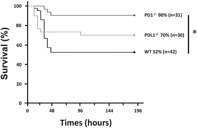
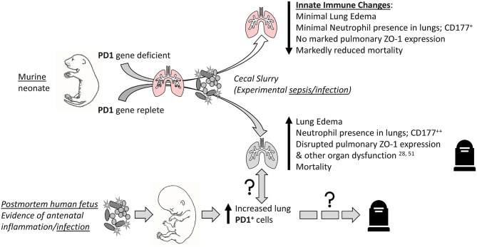
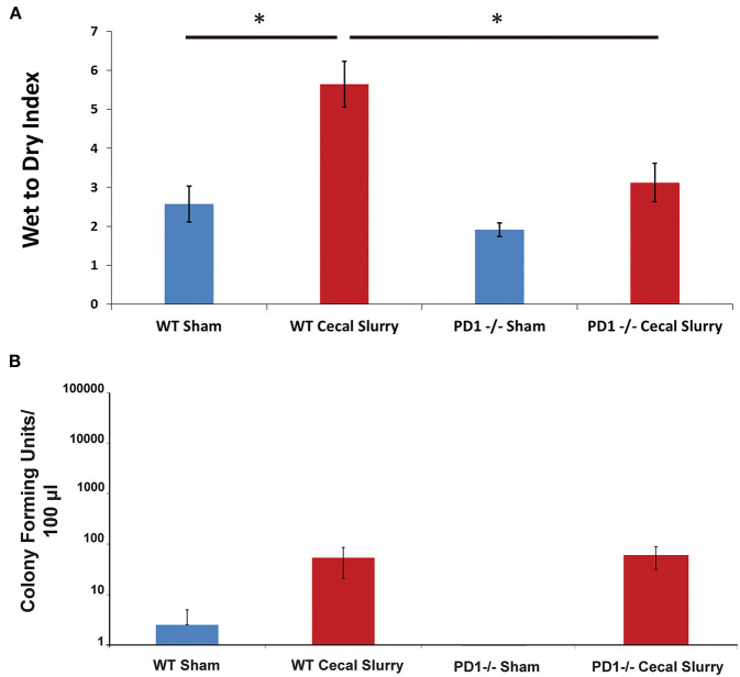
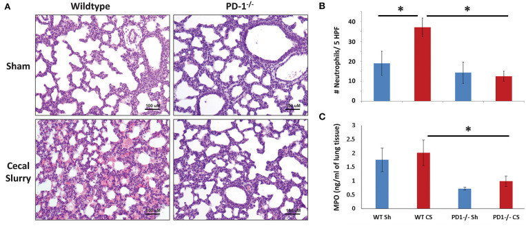
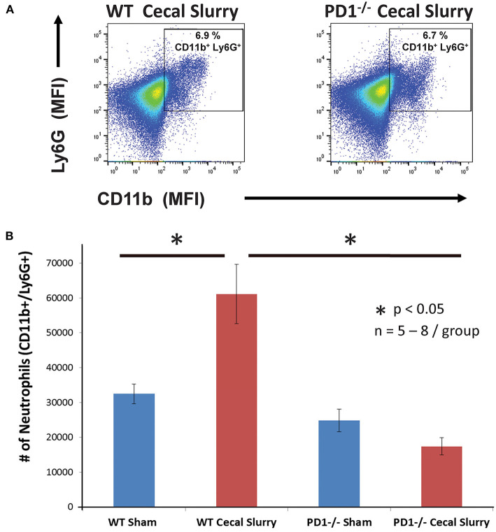
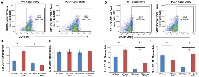
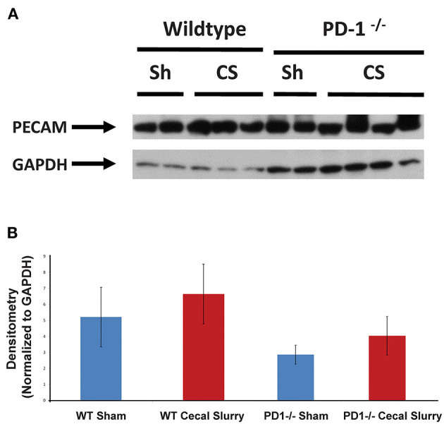
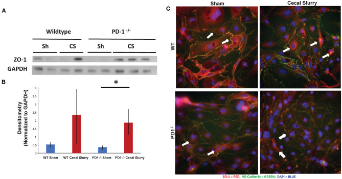
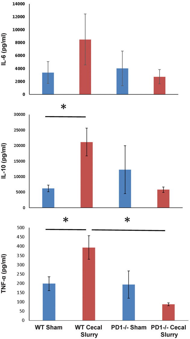
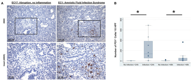
Morbidity and mortality associated with neonatal sepsis remains a healthcare crisis. PD1 neonatal mice endured experimental sepsis, in the form of cecal slurry (CS), and showed improved rates of survival compared to wildtype (WT) counterparts. End-organ injury, particularly of the lung, contributes to the devastation set forth by neonatal sepsis. PDL1 neonatal mice, in contrast to PD1 neonatal mice did not have a significant improvement in survival after CS. Because of this, we focused subsequent studies on the impact of PD1 gene deficiency on lung injury. Here, we observed that at 24 h post-CS (but not at 4 or 12 h) there was a marked increase in pulmonary edema (PE), neutrophil influx, myeloperoxidase (MPO) levels, and cytokine expression sham (Sh) WT mice. Regarding pulmonary endothelial cell (EC) adhesion molecule expression, we observed that Zona occludens-1 (ZO-1) within the cell shifted from a membranous location to a peri-nuclear location after CS in WT murine cultured ECs at 24hrs, but remained membranous among PD1 lungs. To expand the scope of this inquiry, we investigated human neonatal lung tissue. We observed that the lungs of human newborns exposed to intrauterine infection had significantly higher numbers of PD1 cells compared to specimens who died from non-infectious causes. Together, these data suggest that PD1/PDL1, a pathway typically thought to govern adaptive immune processes in adult animals, can modulate the largely innate neonatal pulmonary immune response to experimental septic insult. The potential future significance of this area of study includes that PD1/PDL1 checkpoint proteins may be viable therapeutic targets in the septic neonate.
与新生儿败血症相关的发病率和死亡率仍然是一个医疗保健危机。PD1 新生小鼠经历了实验性败血症,形式为盲肠浆(CS),与野生型(WT)相比,生存率提高。终末器官损伤,特别是肺部损伤,是新生儿败血症造成的破坏的原因。与 PD1 新生小鼠不同,PDL1 新生小鼠在 CS 后生存率没有显著提高。因此,我们将后续研究集中在 PD1 基因缺失对肺损伤的影响上。在这里,我们观察到在 CS 后 24 小时(但不在 4 或 12 小时),肺水肿(PE)、中性粒细胞浸润、髓过氧化物酶(MPO)水平和细胞因子表达明显增加,而在 sham(Sh)WT 小鼠中则没有。关于肺内皮细胞(EC)粘附分子的表达,我们观察到在 CS 后 24 小时,WT 鼠培养的 EC 中 ZO-1 从细胞膜位置转移到核周位置,但在 PD1 肺中仍保持细胞膜位置。为了扩大这一研究范围,我们研究了人新生儿肺组织。我们观察到,与死于非感染性原因的标本相比,暴露于宫内感染的人新生儿肺中有明显更多的 PD1 细胞。这些数据表明,PD1/PDL1 是一种通常被认为在成年动物中调节适应性免疫过程的途径,可以调节新生儿对实验性败血症损伤的主要固有肺免疫反应。这一研究领域的潜在未来意义包括 PD1/PDL1 检查点蛋白可能是败血症新生儿可行的治疗靶点。