Laboratorio de Fisiología Animal, Departamento de Biología Celular, Genética y Fisiología, Facultad de Ciencias, Universidad de Málaga, Instituto de Investigación Biomédica de Málaga-IBIMA, Campus de Teatinos, 29071, Málaga, Spain.
Fluids Barriers CNS. 2021 Mar 23;18(1):15. doi: 10.1186/s12987-021-00249-0.
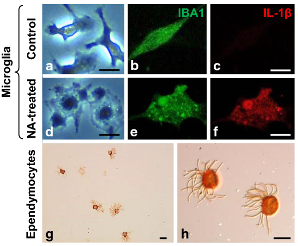
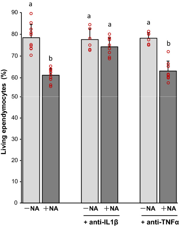
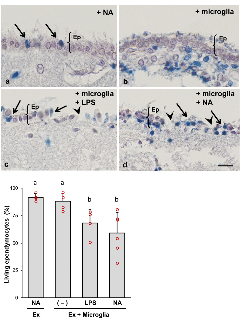
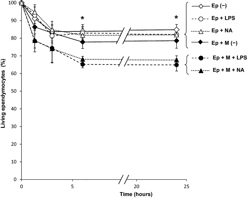
The administration of microbial neuraminidase into the brain ventricular cavities of rodents represents a model of acute aseptic neuroinflammation. Ependymal cell death and hydrocephalus are unique features of this model. Here we demonstrate that activated microglia participates in ependymal cell death. Co-cultures of pure microglia with ependymal cells (both obtained from rats) were performed, and neuraminidase or lipopolysaccharide were used to activate microglia. Ependymal cell viability was unaltered in the absence of microglia or inflammatory stimulus (neuraminidase or lipopolysaccharide). The constitutive expression by ependymal cells of receptors for cytokines released by activated microglia, such as IL-1β, was demonstrated by qPCR. Besides, neuraminidase induced the overexpression of both receptors in ventricular wall explants. Finally, ependymal viability was evaluated in the presence of functional blocking antibodies against IL-1β and TNFα. In the co-culture setting, an IL-1β blocking antibody prevented ependymal cell death, while TNFα antibody did not. These results suggest that activated microglia are involved in the ependymal damage that occurs after the administration of neuraminidase in the ventricular cavities, and points to IL-1β as possible mediator of such effect. The relevance of these results lies in the fact that brain infections caused by neuraminidase-bearing pathogens are frequently associated to ependymal death and hydrocephalus.
将微生物神经氨酸酶注入啮齿动物的脑室内腔代表了一种急性无菌性神经炎症模型。室管膜细胞死亡和脑积水是该模型的独特特征。在这里,我们证明激活的小胶质细胞参与了室管膜细胞的死亡。进行了纯小胶质细胞与室管膜细胞(均来自大鼠)的共培养,并使用神经氨酸酶或脂多糖激活小胶质细胞。在没有小胶质细胞或炎症刺激物(神经氨酸酶或脂多糖)的情况下,室管膜细胞的活力没有改变。通过 qPCR 证明了室管膜细胞表达由激活的小胶质细胞释放的细胞因子(如 IL-1β)的受体。此外,神经氨酸酶诱导了室壁外植体中这两种受体的过表达。最后,在存在针对 IL-1β 和 TNFα 的功能阻断抗体的情况下评估了室管膜细胞的活力。在共培养设置中,IL-1β 阻断抗体可防止室管膜细胞死亡,而 TNFα 抗体则不能。这些结果表明,激活的小胶质细胞参与了神经氨酸酶在脑室腔中给药后发生的室管膜损伤,并且表明 IL-1β 可能是这种作用的介导物。这些结果的相关性在于,携带神经氨酸酶的病原体引起的脑感染常与室管膜细胞死亡和脑积水有关。