Li Ting, Zhao Jin, Xie Wenguang, Yuan Wanru, Guo Jing, Pang Shengru, Gan Wen-Biao, Gómez-Nicola Diego, Zhang Shengxiang
Gansu Key Laboratory of Biomonitoring and Bioremediation for Environmental Pollution, School of Life Sciences, Lanzhou University, No. 222 South Tianshui Road, Lanzhou, Gansu, 730000, People's Republic of China.
Molecular Neurobiology Program, The Kimmel Center for Biology and Medicine of the Skirball Institute, Department of Neuroscience and Physiology, New York University School of Medicine, New York, NY, 10016, USA.
J Neuroinflammation. 2021 Mar 23;18(1):81. doi: 10.1186/s12974-021-02127-w.
Ischemia can induce rapid activation of microglia in the brain. As key immunocompetent cells, reactive microglia play an important role in pathological development of ischemic stroke. However, the role of activated microglia during the development of ischemia remains controversial. Thus, we aimed to investigate the function of reactive microglia in the early stage of ischemic stroke.
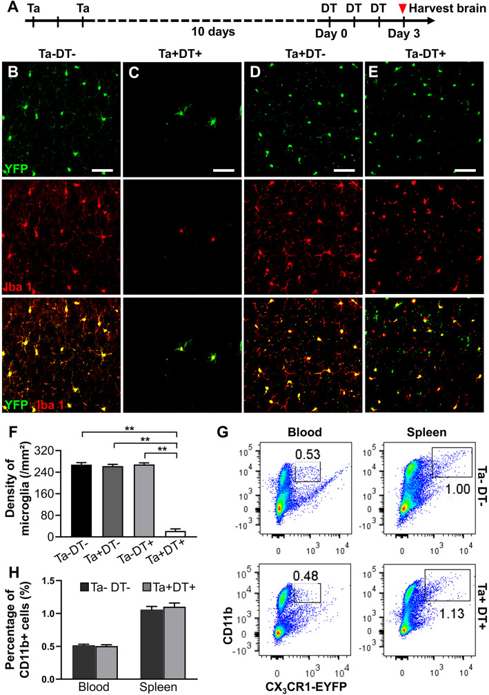
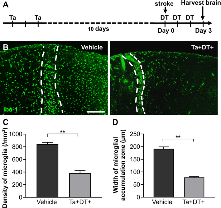
A Rose Bengal photothrombosis model was applied to induce targeted ischemic stroke in mice. CX3CR1:R26 mice were used to specifically deplete resident microglia through intragastric administration of tamoxifen (Ta) and intraperitoneal injection of diphtheria toxin (DT). At day 3 after ischemic stroke, behavioral tests were performed. After that, mouse brains were collected for further histological analysis and detection of mRNA expression of inflammatory factors.
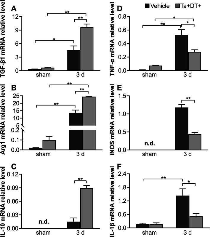
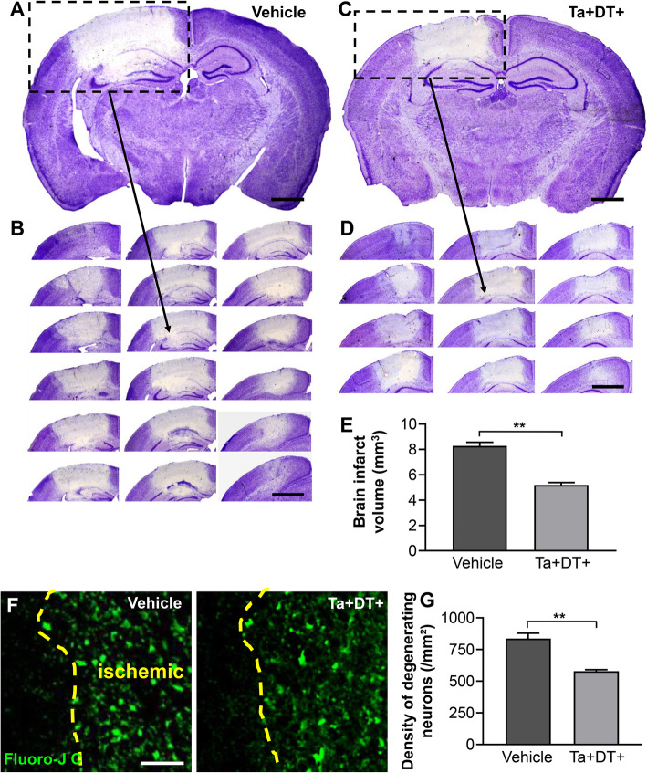
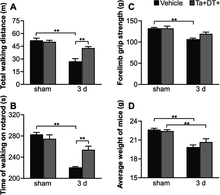
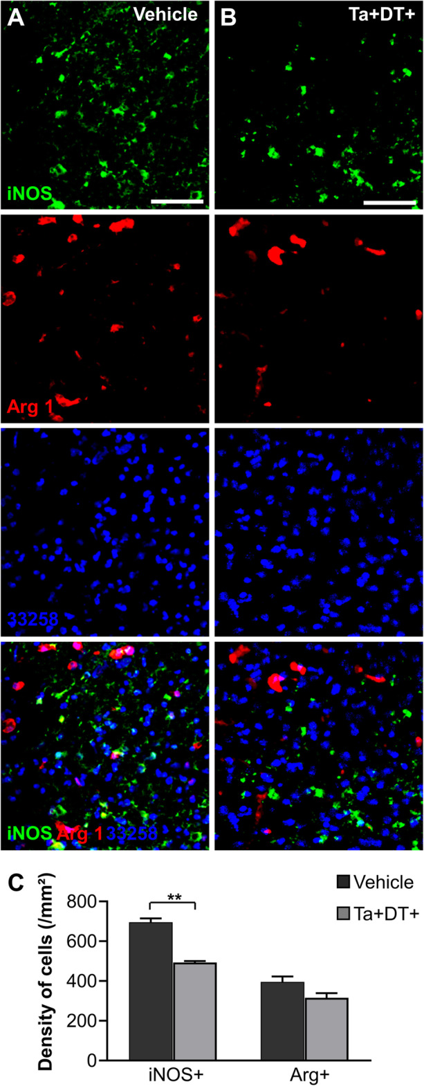
The results showed that specific depletion of microglia resulted in a significant decrease in ischemic infarct volume and improved performance in motor ability 3 days after stroke. Microglial depletion caused a remarkable reduction in the densities of degenerating neurons and inducible nitric oxide synthase positive (iNOS) cells. Importantly, depleting microglia induced a significant increase in the mRNA expression level of anti-inflammatory factors TGF-β1, Arg1, IL-10, IL-4, and Ym1 as well as a significant decline of pro-inflammatory factors TNF-α, iNOS, and IL-1β 3 days after stroke.
These results suggest that activated microglia is an important modulator of the brain's inflammatory response in stroke, contributing to neurological deficit and infarct expansion. Modulation of the inflammatory response through the elimination of microglia at a precise time point may be a promising therapeutic approach for the treatment of cerebral ischemia.
缺血可诱导脑内小胶质细胞快速激活。作为关键的免疫活性细胞,反应性小胶质细胞在缺血性脑卒中的病理发展过程中发挥重要作用。然而,激活的小胶质细胞在缺血发展过程中的作用仍存在争议。因此,我们旨在研究反应性小胶质细胞在缺血性脑卒中早期的功能。
应用孟加拉玫瑰红光血栓形成模型诱导小鼠发生靶向性缺血性脑卒中。利用CX3CR1:R26小鼠,通过胃内给予他莫昔芬(Ta)和腹腔注射白喉毒素(DT)特异性清除驻留小胶质细胞。在缺血性脑卒中后第3天进行行为测试。之后,收集小鼠大脑进行进一步的组织学分析和炎症因子mRNA表达检测。
结果显示,小胶质细胞的特异性清除导致缺血梗死体积显著减小,且在脑卒中后3天运动能力表现得到改善。小胶质细胞清除导致变性神经元和诱导型一氧化氮合酶阳性(iNOS)细胞密度显著降低。重要的是,在脑卒中后3天,清除小胶质细胞导致抗炎因子TGF-β1、Arg1、IL-10、IL-4和Ym1的mRNA表达水平显著升高,同时促炎因子TNF-α、iNOS和IL-1β显著下降。
这些结果表明,激活的小胶质细胞是脑卒中时大脑炎症反应的重要调节因子,促成神经功能缺损和梗死灶扩大。在精确的时间点通过清除小胶质细胞来调节炎症反应可能是治疗脑缺血的一种有前景的治疗方法。