Dolliver Wojciech R, Diaz Alejandro A
Division of Pulmonary and Critical Care Medicine, Brigham and Women's Hospital, Boston, MA, USA.
Barc Respir Netw Rev. 2020 May-Dec;6(2):128-143. doi: 10.23866/brnrev:2019-0023.
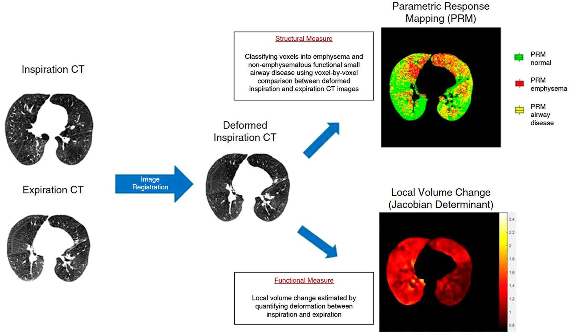
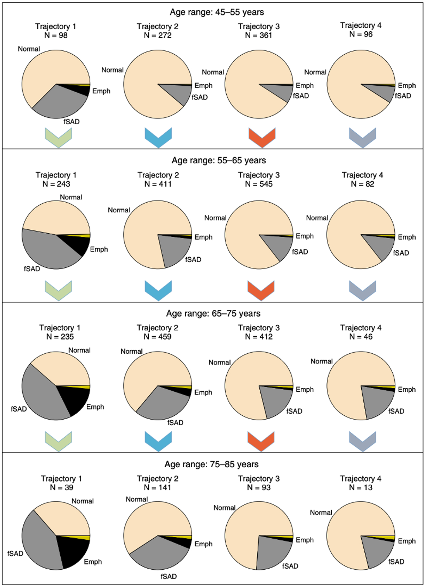
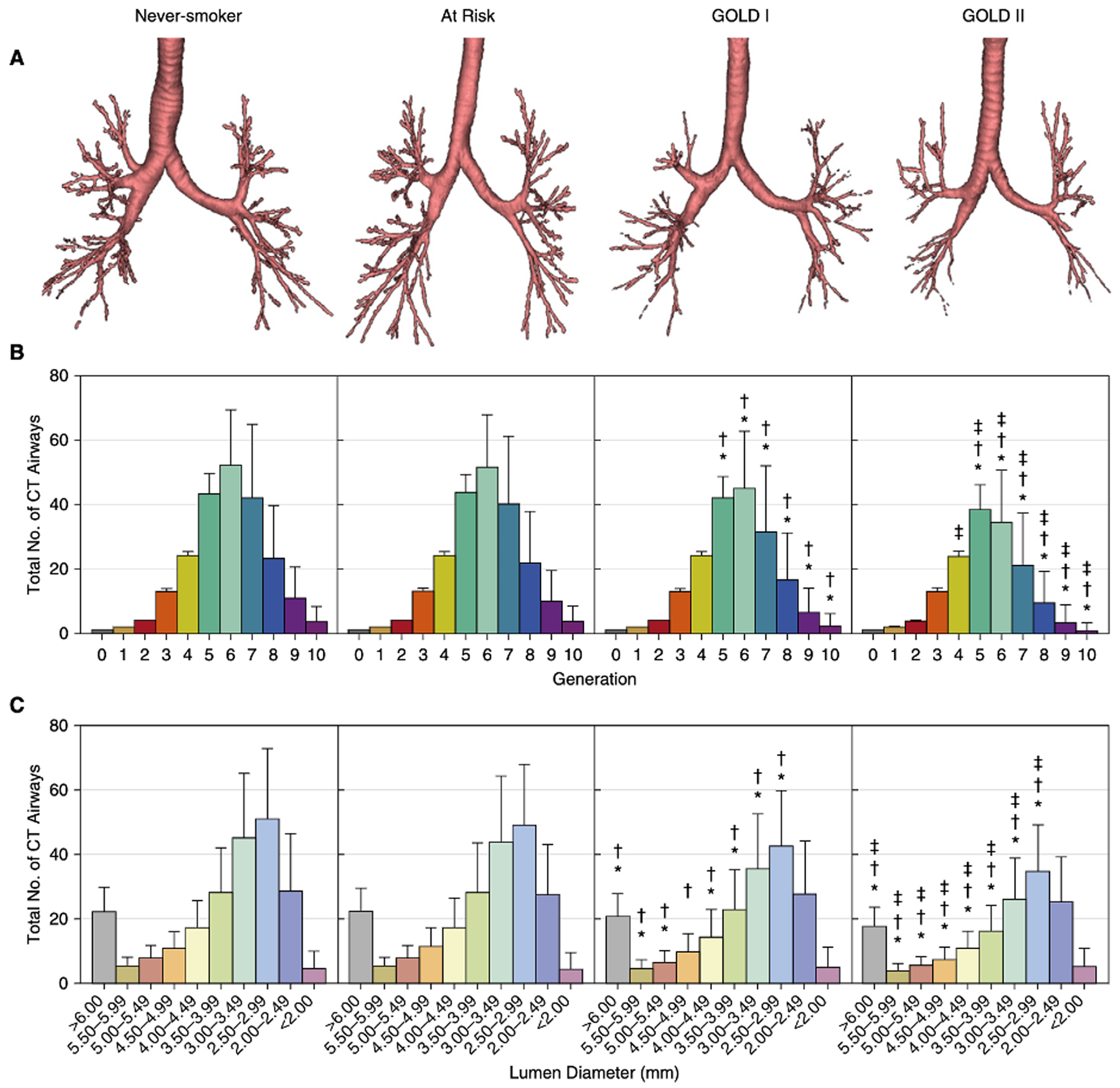
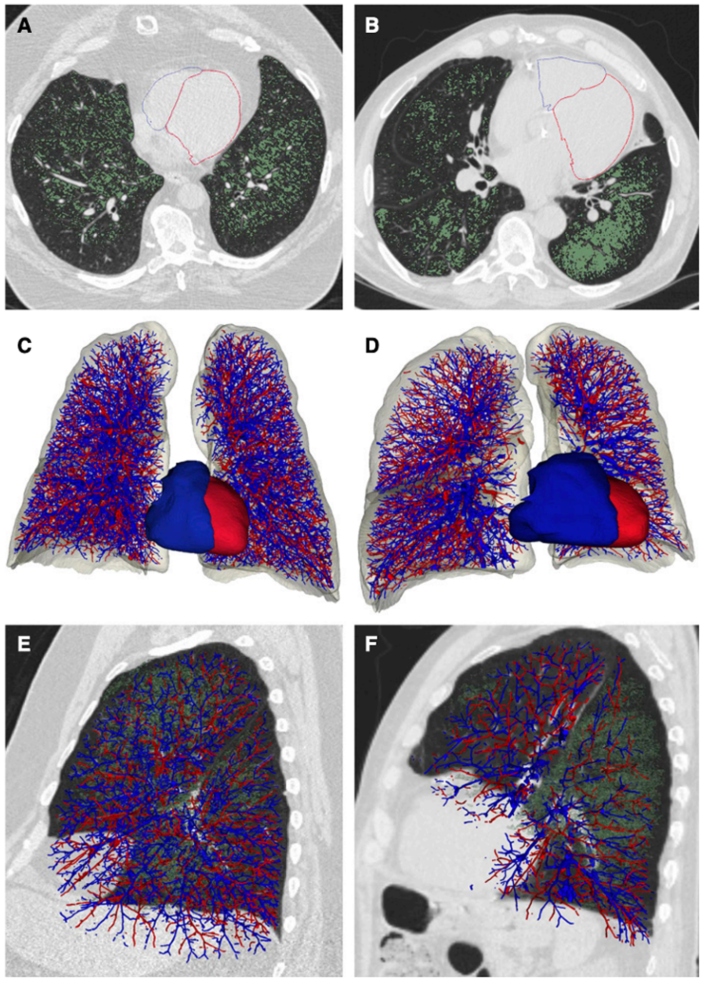
Chest computed tomography (CT) imaging is a useful tool that provides information regarding lung structure. Imaging has contributed to a better understanding of COPD, allowing for the detection of early structural changes and the quantification of extra-pulmonary structures. Novel CT imaging techniques have provided insight into the progression of the main COPD subtypes, such as emphysema and small airway disease. This article serves as a review of new information relevant to COPD imaging. CT abnormalities, such as emphysema and loss of airways, are present even in smokers who do not meet the criteria for COPD and in those with mild-to-moderate disease. Subjects with mild-to-moderate COPD, with the highest loss of airways, also experience the highest decline in lung function. Extra-pulmonary manifestations of COPD, such as right ventricle enlargement and low muscle mass measured on CT, are associated with increased risk for all-cause mortality. CT longitudinal data has also given insight into the progression of COPD. Mechanically affected areas of lung parenchyma adjacent to emphysematous areas are associated with a greater decline in FEV. Subjects with the greatest percentage of small airway disease, as measured on matched inspiratory-expiratory CT scan, also present with the greatest decline in lung function.
胸部计算机断层扫描(CT)成像是一种有用的工具,可提供有关肺结构的信息。成像有助于更好地理解慢性阻塞性肺疾病(COPD),能够检测早期结构变化并对肺外结构进行量化。新型CT成像技术为深入了解COPD的主要亚型(如肺气肿和小气道疾病)的进展提供了线索。本文旨在综述与COPD成像相关的新信息。即使在不符合COPD标准的吸烟者以及轻度至中度疾病患者中,也存在诸如肺气肿和气道缺失等CT异常。气道损失最高的轻度至中度COPD患者,其肺功能下降也最为明显。COPD的肺外表现,如CT测量显示的右心室扩大和肌肉量减少,与全因死亡率增加相关。CT纵向数据也为了解COPD的进展提供了线索。与肺气肿区域相邻的受机械影响的肺实质区域,其第一秒用力呼气容积(FEV)下降幅度更大。在匹配的吸气-呼气CT扫描中测量的小气道疾病比例最高的患者,其肺功能下降也最为明显。