Poly Tahmina Nasrin, Islam Md Mohaimenul, Li Yu-Chuan Jack, Alsinglawi Belal, Hsu Min-Huei, Jian Wen Shan, Yang Hsuan-Chia
Graduate Institute of Biomedical Informatics, College of Medical Science and Technology, Taipei Medical University, Taipei, Taiwan.
International Center for Health Information Technology, Taipei Medical University, Taipei, Taiwan.
JMIR Med Inform. 2021 Apr 29;9(4):e21394. doi: 10.2196/21394.
The COVID-19 outbreak has spread rapidly and hospitals are overwhelmed with COVID-19 patients. While analysis of nasal and throat swabs from patients is the main way to detect COVID-19, analyzing chest images could offer an alternative method to hospitals, where health care personnel and testing kits are scarce. Deep learning (DL), in particular, has shown impressive levels of performance when analyzing medical images, including those related to COVID-19 pneumonia.
The goal of this study was to perform a systematic review with a meta-analysis of relevant studies to quantify the performance of DL algorithms in the automatic stratification of COVID-19 patients using chest images.
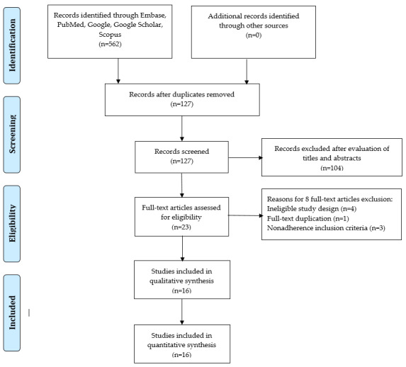
A search strategy for use in PubMed, Scopus, Google Scholar, and Web of Science was developed, where we searched for articles published between January 1 and April 25, 2020. We used the key terms "COVID-19," or "coronavirus," or "SARS-CoV-2," or "novel corona," or "2019-ncov," and "deep learning," or "artificial intelligence," or "automatic detection." Two authors independently extracted data on study characteristics, methods, risk of bias, and outcomes. Any disagreement between them was resolved by consensus.
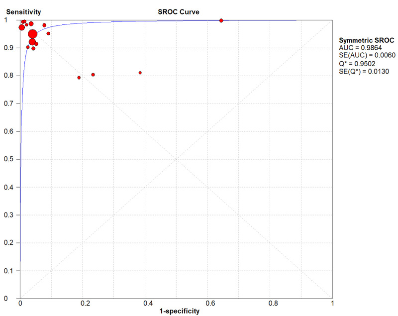
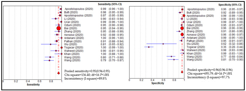
A total of 16 studies were included in the meta-analysis, which included 5896 chest images from COVID-19 patients. The pooled sensitivity and specificity of the DL models in detecting COVID-19 were 0.95 (95% CI 0.94-0.95) and 0.96 (95% CI 0.96-0.97), respectively, with an area under the receiver operating characteristic curve of 0.98. The positive likelihood, negative likelihood, and diagnostic odds ratio were 19.02 (95% CI 12.83-28.19), 0.06 (95% CI 0.04-0.10), and 368.07 (95% CI 162.30-834.75), respectively. The pooled sensitivity and specificity for distinguishing other types of pneumonia from COVID-19 were 0.93 (95% CI 0.92-0.94) and 0.95 (95% CI 0.94-0.95), respectively. The performance of radiologists in detecting COVID-19 was lower than that of the DL models; however, the performance of junior radiologists was improved when they used DL-based prediction tools.
Our study findings show that DL models have immense potential in accurately stratifying COVID-19 patients and in correctly differentiating them from patients with other types of pneumonia and normal patients. Implementation of DL-based tools can assist radiologists in correctly and quickly detecting COVID-19 and, consequently, in combating the COVID-19 pandemic.
新型冠状病毒肺炎(COVID-19)疫情迅速蔓延,医院里COVID-19患者数量激增,不堪重负。虽然对患者的鼻咽拭子分析是检测COVID-19的主要方法,但对于医疗人员和检测试剂盒稀缺的医院而言,分析胸部影像可能提供一种替代方法。尤其是深度学习(DL)在分析医学影像(包括与COVID-19肺炎相关的影像)时表现出了令人瞩目的性能水平。
本研究的目的是进行一项系统评价,并对相关研究进行荟萃分析,以量化DL算法在使用胸部影像对COVID-19患者进行自动分层方面的性能。
制定了在PubMed、Scopus、谷歌学术和科学网中使用的检索策略,检索2020年1月1日至4月25日期间发表的文章。我们使用了关键词“COVID-19”、“冠状病毒”、“严重急性呼吸综合征冠状病毒2(SARS-CoV-2)”、“新型冠状病毒”、“2019新型冠状病毒(2019-ncov)”以及“深度学习”、“人工智能”、“自动检测”。两名作者独立提取关于研究特征、方法、偏倚风险和结果的数据。他们之间的任何分歧都通过协商解决。
荟萃分析共纳入16项研究,其中包括来自COVID-19患者的5896张胸部影像。DL模型检测COVID-19的合并敏感度和特异度分别为0.95(95%CI 0.94 - 0.95)和0.96(95%CI 0.96 - 0.97),受试者工作特征曲线下面积为0.98。阳性似然比、阴性似然比和诊断比值比分别为19.02(95%CI 12.83 - 28.19)、0.06(95%CI 0.04 - 0.10)和368.07(95%CI 162.30 - 834.75)。区分其他类型肺炎与COVID-19的合并敏感度和特异度分别为0.93(95%CI 0.92 - 0.94)和0.95(95%CI 0.94 - 0.95)。放射科医生检测COVID-19的性能低于DL模型;然而,初级放射科医生使用基于DL的预测工具时,其性能有所提高。
我们的研究结果表明,DL模型在准确分层COVID-19患者以及将其与其他类型肺炎患者和正常患者正确区分方面具有巨大潜力。基于DL的工具的应用可以帮助放射科医生正确、快速地检测COVID-19,从而对抗COVID-19疫情。