Lo Cody, Vuong Laurel N, Micieli Jonathan A
Faculty of Medicine, University of British Columbia, Vancouver, BC, Canada.
The New England Eye Center, Tufts Medical Center, Boston, MA, USA.
Taiwan J Ophthalmol. 2021 Jan 20;11(1):3-15. doi: 10.4103/tjo.tjo_76_20. eCollection 2021 Jan-Mar.
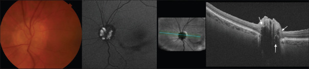
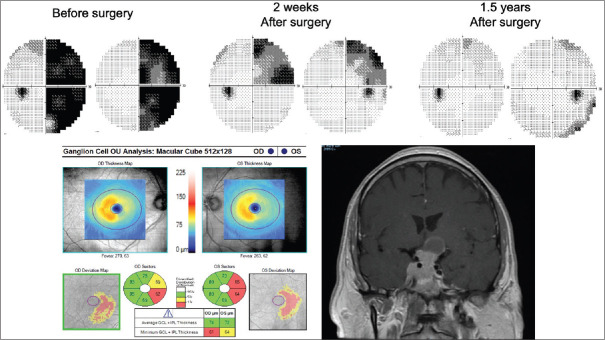
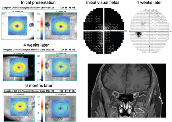
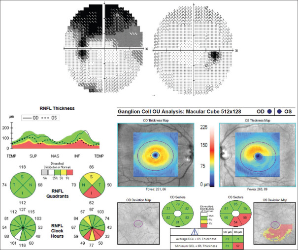
Optical coherence tomography (OCT) is a noninvasive imaging technique used to qualitatively and quantitatively analyze various layers of the retina. OCT of the retinal nerve fiber layer (RNFL) and ganglion cell-inner plexiform layer (GCIPL) is particularly useful in neuro-ophthalmology for the evaluation of patients with optic neuropathies and retrochiasmal visual pathway disorders. OCT allows for an objective quantification of edema and atrophy of the RNFL and GCIPL, which may be evident before obvious clinical signs and visual dysfunction develop. Enhanced depth imaging OCT allows for visualization of deep structures of the optic nerve and has emerged as the gold standard for the detection of optic disc drusen. In the evaluation of compressive optic neuropathies, OCT RNFL and GCIPL thicknesses have been established as the most important visual prognostic factor. There is increasing evidence that inclusion of OCT as part of the diagnostic criteria for multiple sclerosis (MS) increases its sensitivity. Moreover, OCT of the RNFL and GCIPL may be helpful in the early detection and monitoring the treatment of conditions such as MS and Alzheimer's disease. OCT is an important aspect of the neuro-ophthalmologic assessment and its use is likely to increase moving forward.
光学相干断层扫描(OCT)是一种非侵入性成像技术,用于定性和定量分析视网膜的各层结构。视网膜神经纤维层(RNFL)和神经节细胞-内丛状层(GCIPL)的OCT在神经眼科中对于评估视神经病变和视交叉后视觉通路障碍患者特别有用。OCT能够客观量化RNFL和GCIPL的水肿和萎缩情况,这些情况在明显的临床体征和视觉功能障碍出现之前可能就已显现。增强深度成像OCT能够对视神经的深部结构进行可视化,已成为检测视盘小疣的金标准。在评估压迫性视神经病变时,OCT测量的RNFL和GCIPL厚度已被确立为最重要的视觉预后因素。越来越多的证据表明,将OCT纳入多发性硬化症(MS)的诊断标准可提高其敏感性。此外,RNFL和GCIPL的OCT可能有助于早期检测和监测MS及阿尔茨海默病等疾病的治疗情况。OCT是神经眼科评估的一个重要方面,并且其应用可能会在未来不断增加。