• 文献检索
  • 文档翻译
  • 深度研究
  • 学术资讯
  • Suppr Zotero 插件Zotero 插件
  • 邀请有礼
  • 套餐&价格
  • 历史记录
应用&插件
Suppr Zotero 插件Zotero 插件浏览器插件Mac 客户端Windows 客户端微信小程序
定价
高级版会员购买积分包购买API积分包
服务
文献检索文档翻译深度研究API 文档MCP 服务
关于我们
关于 Suppr公司介绍联系我们用户协议隐私条款
关注我们

Suppr 超能文献

核心技术专利:CN118964589B侵权必究
粤ICP备2023148730 号-1Suppr @ 2026

文献检索

告别复杂PubMed语法,用中文像聊天一样搜索,搜遍4000万医学文献。AI智能推荐,让科研检索更轻松。

立即免费搜索

文件翻译

保留排版,准确专业,支持PDF/Word/PPT等文件格式,支持 12+语言互译。

免费翻译文档

深度研究

AI帮你快速写综述,25分钟生成高质量综述,智能提取关键信息,辅助科研写作。

立即免费体验

光学相干断层扫描血管造影术在预测经鼻内镜垂体手术后解剖和功能恢复中的作用:一项为期 1 年的纵向研究。

The role of OCT- angiography in predicting anatomical and functional recovery after endoscopic endonasal pituitary surgery: A 1-year longitudinal study.

机构信息

Public Health Department, Università degli Studi di Napoli "Federico II", Naples, Italy.

Division of Neurosurgery, Department of Neurosciences, Reproductive Sciences and Odontostomatological Sciences, Università degli Studi di Napoli "Federico II", Naples, Italy.

出版信息

PLoS One. 2021 Dec 2;16(12):e0260029. doi: 10.1371/journal.pone.0260029. eCollection 2021.

DOI:10.1371/journal.pone.0260029
PMID:34855775
原文链接:https://pmc.ncbi.nlm.nih.gov/articles/PMC8638874/
Abstract

PURPOSE

The purpose of this study was to investigate the changes in structural spectral-domain optical coherence tomography (SD-OCT), OCT Angiography (OCTA) parameters, and visual acuity, 1 year after endoscopic endonasal approach for the removal of an intra-suprasellar pituitary adenoma compressing optic chiasm and compare outcomes with 48 hours postoperative data.

METHODS

Sixteen eyes of eight patients (4 males, 4 females, mean age 52 ± 11 years) were enrolled in this prospective study. The primary outcome was to evaluate the changes over time before and after surgery, analyzing the Best Corrected Visual Acuity (BCVA), Ganglion Cell Complex (GCC), Retinal Nerve Fiber Layer (RNFL) thicknesses, the retinal vessel density (VD) of Superficial Capillary Plexus (SCP), Deep Capillary Plexus (DCP), Radial Peripapillary Capillary (RPC) and the Foveal Avascular Zone (FAZ). The secondary outcome was to identify potential biomarkers that could predict visual acuity changes after 1-year follow-up.

RESULTS

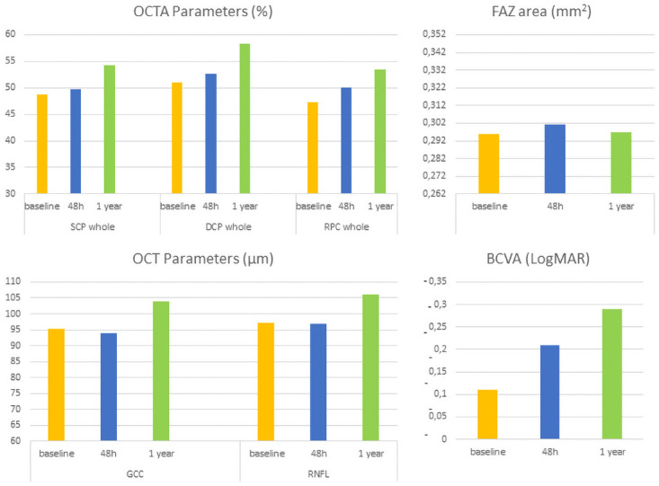
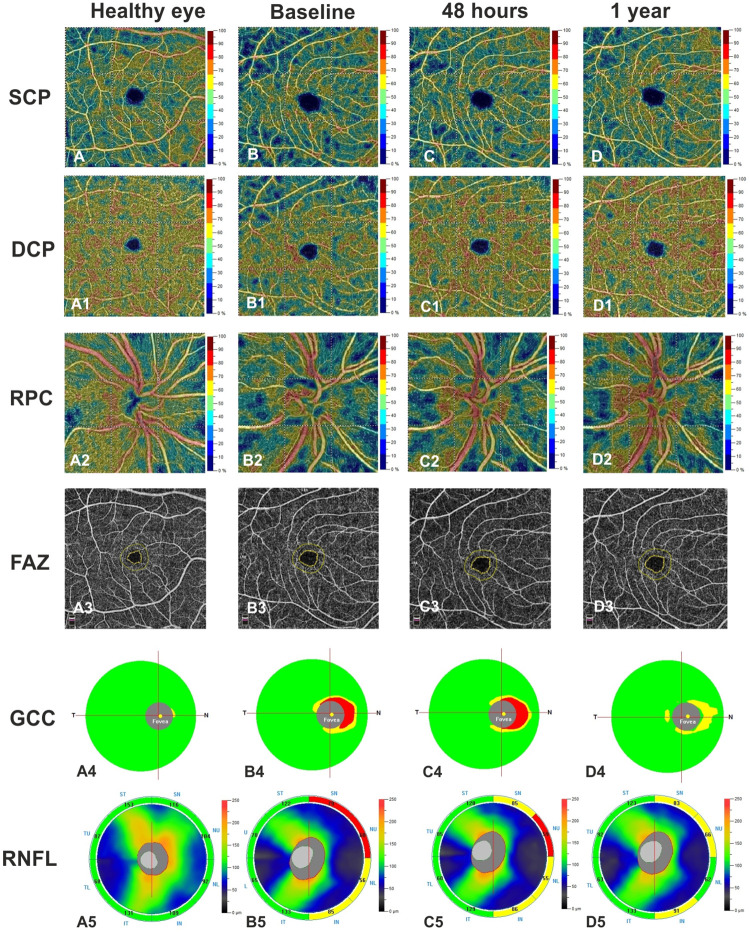
When comparing SD-OCT and OCTA measurements obtained after 1 year with those observed 48 hours after surgery, GCC and RNFL were significantly improved. After a significant reduction at 48 hours, GCC thickness showed a significant increase at 1 year after surgery (p = 0.007), while a significant restoration of RNFL thickness was found at 1 year (p = 0.005), as well as the VD of SCP, DCP, and RPC values. FAZ area did not change over time. BCVA significantly improved at each time after surgery (p = 0.037, p = 0.013). A statistically significant correlation was found between the preoperative BCVA, VD of SCP, DCP, RPC, and the postoperative BCVA at 1 year (p = 0.017, p = 0.029, p = 0.031, p = 0.023).

CONCLUSION

SD-OCT and OCTA provide helpful information to identify the retinal structural and vascular improvements 1 year after surgery. OCTA parameters could serve as potential predictive markers for visual acuity recovery at long-term follow-up.

摘要

目的

本研究旨在探讨内镜经鼻蝶入路切除对视神经交叉受压的鞍上垂体腺瘤患者术后 1 年结构谱域光相干断层扫描(SD-OCT)、OCT 血管造影(OCTA)参数和视力的变化,并与术后 48 小时的数据进行比较。

方法

本前瞻性研究纳入了 8 例患者(4 名男性,4 名女性;平均年龄 52±11 岁)的 16 只眼。主要结局是评估手术前后随时间的变化,分析最佳矫正视力(BCVA)、节细胞复合体(GCC)、视网膜神经纤维层(RNFL)厚度、浅层毛细血管丛(SCP)、深层毛细血管丛(DCP)、放射状神经纤维层(RPC)和中心凹无血管区(FAZ)的视网膜血管密度(VD)。次要结局是确定可能预测术后 1 年视力变化的生物标志物。

结果

与术后 48 小时相比,术后 1 年时 SD-OCT 和 OCTA 测量值显示 GCC 和 RNFL 显著改善。GCC 厚度在术后 48 小时显著降低后,在术后 1 年时显著增加(p=0.007),而 RNFL 厚度在术后 1 年时显著恢复(p=0.005),SCP、DCP 和 RPC 的 VD 值也显著增加。FAZ 面积无随时间变化。术后每次随访 BCVA 均显著改善(p=0.037,p=0.013)。术前 BCVA、SCP、DCP、RPC 的 VD 与术后 1 年时的 BCVA 之间存在显著相关性(p=0.017,p=0.029,p=0.031,p=0.023)。

结论

SD-OCT 和 OCTA 提供了有价值的信息,可识别术后 1 年时视网膜结构和血管的改善。OCTA 参数可作为长期随访中视力恢复的潜在预测标志物。

https://cdn.ncbi.nlm.nih.gov/pmc/blobs/1359/8638874/11125790dd38/pone.0260029.g002.jpg
https://cdn.ncbi.nlm.nih.gov/pmc/blobs/1359/8638874/1bddf0bfdaeb/pone.0260029.g001.jpg
https://cdn.ncbi.nlm.nih.gov/pmc/blobs/1359/8638874/11125790dd38/pone.0260029.g002.jpg
https://cdn.ncbi.nlm.nih.gov/pmc/blobs/1359/8638874/1bddf0bfdaeb/pone.0260029.g001.jpg
https://cdn.ncbi.nlm.nih.gov/pmc/blobs/1359/8638874/11125790dd38/pone.0260029.g002.jpg

相似文献

1
The role of OCT- angiography in predicting anatomical and functional recovery after endoscopic endonasal pituitary surgery: A 1-year longitudinal study.光学相干断层扫描血管造影术在预测经鼻内镜垂体手术后解剖和功能恢复中的作用:一项为期 1 年的纵向研究。
PLoS One. 2021 Dec 2;16(12):e0260029. doi: 10.1371/journal.pone.0260029. eCollection 2021.
2
Early vascular modifications after endoscopic endonasal pituitary surgery: The role of OCT-angiography.经鼻内镜垂体手术后早期血管改变:OCT 血管造影的作用。
PLoS One. 2020 Oct 29;15(10):e0241295. doi: 10.1371/journal.pone.0241295. eCollection 2020.
3
[Correlation of capillary plexus with visual acuity in idiopathic macular epiretinal membrane eyes using optical coherence tomography angiography].[利用光学相干断层扫描血管造影术研究特发性黄斑视网膜前膜眼中毛细血管丛与视力的相关性]
Zhonghua Yan Ke Za Zhi. 2019 Oct 11;55(10):757-762. doi: 10.3760/cma.j.issn.0412-4081.2019.10.006.
4
Association of Optical Coherence Tomography and Optical Coherence Tomography Angiography Retinal Features With Visual Function in Older Adults.老年人光学相干断层扫描和光学相干断层扫描血管造影视网膜特征与视觉功能的关系。
JAMA Ophthalmol. 2022 Aug 1;140(8):809-817. doi: 10.1001/jamaophthalmol.2022.2099.
5
Retinal Vascular Features in Ocular Blunt Trauma by Optical Coherence Tomography Angiography.光学相干断层扫描血管造影术观察钝性眼外伤中的视网膜血管特征
J Clin Med. 2020 Oct 16;9(10):3329. doi: 10.3390/jcm9103329.
6
Changes in retinal vessel and retinal layer thickness after cross-linking in keratoconus via swept-source OCT angiography.交联治疗圆锥角膜后通过扫频源 OCT 血管造影观察视网膜血管和视网膜层厚度的变化。
Graefes Arch Clin Exp Ophthalmol. 2022 Dec;260(12):3919-3925. doi: 10.1007/s00417-022-05749-9. Epub 2022 Jul 6.
7
Peripapillary and macular morpho-vascular changes in patients with genetic or clinical diagnosis of autosomal dominant optic atrophy: a case-control study.常染色体显性遗传性视神经萎缩患者的视乳头周围及黄斑区形态血管变化:一项病例对照研究
Graefes Arch Clin Exp Ophthalmol. 2019 May;257(5):1019-1027. doi: 10.1007/s00417-019-04267-5. Epub 2019 Feb 24.
8
Evaluation of the Optic Disc and Macula in Healthy Children Using Optical Coherence Tomography Angiography.利用光相干断层扫描血管造影评估健康儿童的视盘和黄斑。
Turk J Ophthalmol. 2020 Aug 26;50(4):228-233. doi: 10.4274/tjo.galenos.2020.85282.
9
Optical Coherence Tomography Angiography Features in Post-COVID-19 Pneumonia Patients: A Pilot Study.新冠后肺炎患者的光学相干断层扫描血管造影特征:一项初步研究
Am J Ophthalmol. 2021 Jul;227:182-190. doi: 10.1016/j.ajo.2021.03.015. Epub 2021 Mar 27.
10
Macular capillary plexuses after epiretinal membrane surgery: an optical coherence tomography angiography study.视网膜膜后手术的黄斑毛细血管丛:光学相干断层扫描血管造影研究。
Br J Ophthalmol. 2018 Aug;102(8):1086-1091. doi: 10.1136/bjophthalmol-2017-311188. Epub 2017 Oct 31.

引用本文的文献

1
Multidisciplinary management of pituitary macroadenoma.垂体大腺瘤的多学科管理
World J Methodol. 2025 Sep 20;15(3):97694. doi: 10.5662/wjm.v15.i3.97694.
2
Long-term retinal microvascular alterations after endoscopic endonasal surgery in patients with sellar region tumors.鞍区肿瘤患者经鼻内镜手术后的长期视网膜微血管改变
Int J Ophthalmol. 2025 Aug 18;18(8):1515-1521. doi: 10.18240/ijo.2025.08.13. eCollection 2025.
3
Impact of Oral Citicoline, Antioxidant Vitamins, and Blackcurrant Supplementation on Primary Open-Angle Glaucoma: An OCT and OCTA Study.

本文引用的文献

1
Recent advances and future directions on the use of optical coherence tomography in neuro-ophthalmology.光学相干断层扫描在神经眼科应用中的最新进展与未来方向
Taiwan J Ophthalmol. 2021 Jan 20;11(1):3-15. doi: 10.4103/tjo.tjo_76_20. eCollection 2021 Jan-Mar.
2
Analysis of factors affecting visual field recovery following surgery for pituitary adenoma.分析影响垂体腺瘤手术后视野恢复的因素。
Int Ophthalmol. 2021 Jun;41(6):2019-2026. doi: 10.1007/s10792-021-01757-6. Epub 2021 Feb 24.
3
Early vascular modifications after endoscopic endonasal pituitary surgery: The role of OCT-angiography.
口服胞磷胆碱、抗氧化维生素和黑加仑补充剂对原发性开角型青光眼的影响:一项光学相干断层扫描(OCT)和光学相干断层扫描血管造影(OCTA)研究
Biomedicines. 2025 May 31;13(6):1352. doi: 10.3390/biomedicines13061352.
4
Resistive index of central retinal artery, aortic arterial stiffness and OCTA correlated parameters in the early stage of fabry disease.中央视网膜动脉阻力指数、主动脉动脉僵硬度和 OCTA 相关参数在法布里病早期的相关性。
Sci Rep. 2024 Oct 14;14(1):24047. doi: 10.1038/s41598-024-74146-5.
5
Application of Optical Coherence Tomography Angiography in True and Pseudo-Optic Disc Swelling.光学相干断层扫描血管造影在真性和假性视盘水肿中的应用。
J Ophthalmol. 2024 Sep 30;2024:1164635. doi: 10.1155/2024/1164635. eCollection 2024.
6
Comparative of OCT and OCTA parameters in patients with early chronic angle-closure glaucoma and early pituitary adenoma.比较早期慢性闭角型青光眼和早期垂体腺瘤患者的 OCT 和 OCTA 参数。
Sci Rep. 2024 Sep 13;14(1):21448. doi: 10.1038/s41598-024-71103-0.
7
The Role of Optical Coherence Tomography Angiography in the Evaluation of Chiasmal and Retrochiasmal Compression.光学相干断层扫描血管造影在视交叉及视交叉后压迫评估中的作用
Neuroophthalmology. 2024 Apr 19;48(5):315-327. doi: 10.1080/01658107.2024.2338411. eCollection 2024.
8
Insights into Visual Outcomes and Determinants in Pituitary Adenoma Surgery: A 5-Year Retrospective Cohort Study.垂体腺瘤手术的视觉结果及决定因素分析:一项5年回顾性队列研究
Clin Ophthalmol. 2024 Jun 21;18:1779-1788. doi: 10.2147/OPTH.S463303. eCollection 2024.
9
Predictive visual field outcomes after optic chiasm decompressive surgery by retinal vessels parameters using optical coherence tomography angiography.利用光学相干断层扫描血管造影术通过视网膜血管参数预测视交叉减压术后的视野结果。
Int J Ophthalmol. 2024 Feb 18;17(2):365-373. doi: 10.18240/ijo.2024.02.21. eCollection 2024.
经鼻内镜垂体手术后早期血管改变:OCT 血管造影的作用。
PLoS One. 2020 Oct 29;15(10):e0241295. doi: 10.1371/journal.pone.0241295. eCollection 2020.
4
Visual acuity and its postoperative outcome after transsphenoidal adenoma resection.经蝶窦腺瘤切除术的视力及其术后结果。
Neurosurg Rev. 2021 Aug;44(4):2245-2251. doi: 10.1007/s10143-020-01408-x. Epub 2020 Oct 10.
5
An Analysis of Retinal Nerve Fiber Layer Thickness before and after Pituitary Adenoma Surgery and its Correlation with Visual Acuity.垂体瘤手术后视网膜神经纤维层厚度的分析及其与视力的相关性。
Neurol India. 2020 Mar-Apr;68(2):346-351. doi: 10.4103/0028-3886.280634.
6
Parafoveal and Peripapillary Perfusion Predict Visual Field Recovery in Chiasmal Compression due to Pituitary Tumors.黄斑旁和视乳头周围灌注可预测垂体肿瘤所致视交叉受压患者的视野恢复情况。
J Clin Med. 2020 Mar 4;9(3):697. doi: 10.3390/jcm9030697.
7
Analysis of Optic Chiasmal Compression Caused by Brain Tumors Using Optical Coherence Tomography Angiography.利用光相干断层扫描血管造影术分析脑肿瘤对视交叉的压迫。
Sci Rep. 2020 Feb 7;10(1):2088. doi: 10.1038/s41598-020-59158-1.
8
Retinal microvasculature in pituitary adenoma patients: is optical coherence tomography angiography useful?垂体腺瘤患者的视网膜微血管系统:光学相干断层扫描血管造影术是否有用?
Acta Ophthalmol. 2020 Aug;98(5):e585-e592. doi: 10.1111/aos.14322. Epub 2019 Dec 6.
9
Predicting the early visual outcomes in sellar-suprasellar lesions compressing the chiasm: the role of SD-OCT series of 20 patients operated via endoscopic endonasal approach.预测经内镜经鼻入路手术治疗鞍上-鞍旁病变对视交叉压迫患者的早期视力结局:20 例患者的 SD-OCT 系列检查的作用。
J Neurosurg Sci. 2022 Aug;66(4):362-370. doi: 10.23736/S0390-5616.19.04687-3. Epub 2019 Jul 23.
10
Comparison of methods to quantify macular and peripapillary vessel density in optical coherence tomography angiography.比较光学相干断层扫描血管造影术定量黄斑和视盘周围血管密度的方法。
PLoS One. 2018 Oct 18;13(10):e0205773. doi: 10.1371/journal.pone.0205773. eCollection 2018.