Academic Department of Magnetic Resonance Imaging, University of Sheffield, Sheffield, UK.
Department of Oncology and Human Metabolism, University of Sheffield, Sheffield, UK.
Diabetologia. 2021 Jun;64(6):1412-1421. doi: 10.1007/s00125-021-05416-4. Epub 2021 Mar 25.
AIMS/HYPOTHESIS: The aim of this work was to investigate whether different clinical pain phenotypes of diabetic polyneuropathy (DPN) are distinguished by functional connectivity at rest.
This was an observational, cohort study of 43 individuals with painful DPN, divided into irritable (IR, n = 10) and non-irritable (NIR, n = 33) nociceptor phenotypes using the German Research Network of Neuropathic Pain quantitative sensory testing protocol. In-situ brain MRI included 3D T1-weighted anatomical and 6 min resting-state functional MRI scans. Subgroup differences in resting-state functional connectivity in brain regions involved with somatic (thalamus, primary somatosensory cortex, motor cortex) and non-somatic (insular and anterior cingulate cortices) pain processing were examined. Multidimensional reduction of MRI datasets was performed using a machine-learning approach to classify individuals into each clinical pain phenotype.
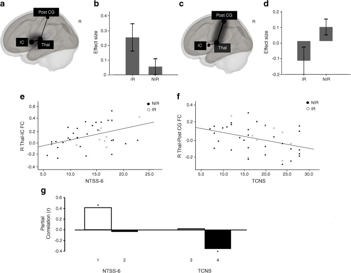
Individuals with the IR nociceptor phenotype had significantly greater thalamic-insular cortex (p false discovery rate [FDR] = 0.03) and reduced thalamus-somatosensory cortex functional connectivity (p-FDR = 0.03). We observed a double dissociation such that self-reported neuropathic pain score was more associated with greater thalamus-insular cortex functional connectivity (r = 0.41; p = 0.01) whereas more severe nerve function deficits were more related to lower thalamus-somatosensory cortex functional connectivity (r = -0.35; p = 0.03). Machine-learning group classification performance to identify individuals with the NIR nociceptor phenotype achieved an accuracy of 0.92 (95% CI 0.08) and sensitivity of 90%.
CONCLUSIONS/INTERPRETATION: This study demonstrates differences in functional connectivity in nociceptive processing brain regions between IR and NIR phenotypes in painful DPN. We also establish proof of concept for the utility of multimodal MRI as a biomarker for painful DPN by using a machine-learning approach to classify individuals into sensory phenotypes.
目的/假设:本研究旨在探究不同的糖尿病周围神经病变(DPN)临床疼痛表型是否可通过静息状态下的功能连接来区分。
这是一项观察性队列研究,纳入 43 例有疼痛症状的 DPN 患者,根据德国神经病学学会神经性疼痛定量感觉测试方案将其分为易激惹(IR,n=10)和非易激惹(NIR,n=33)感觉神经表型。原位脑 MRI 包括 3D T1 加权解剖和 6 分钟静息态功能 MRI 扫描。检查躯体(丘脑、初级躯体感觉皮层、运动皮层)和非躯体(岛叶和前扣带回皮层)疼痛处理相关脑区静息态功能连接的亚组差异。使用机器学习方法对 MRI 数据集进行多维降维,以将个体分类为每个临床疼痛表型。
IR 感觉神经表型个体的丘脑-岛叶皮层功能连接明显增加(假发现率 [FDR] p=0.03),丘脑-躯体感觉皮层功能连接减少(p-FDR=0.03)。我们观察到一种双重分离现象,即自我报告的神经性疼痛评分与更大的丘脑-岛叶皮层功能连接相关性更强(r=0.41;p=0.01),而更严重的神经功能缺陷与更低的丘脑-躯体感觉皮层功能连接相关性更强(r=-0.35;p=0.03)。用于识别 NIR 感觉神经表型个体的机器学习组分类性能达到 0.92(95%置信区间 0.08)的准确率和 90%的敏感度。
本研究表明,在有疼痛症状的 DPN 中,IR 和 NIR 表型之间的痛觉处理脑区的功能连接存在差异。我们还通过使用机器学习方法对个体进行感觉表型分类,为多模态 MRI 作为疼痛性 DPN 的生物标志物的实用性提供了概念验证。