• 文献检索
  • 文档翻译
  • 深度研究
  • 学术资讯
  • Suppr Zotero 插件Zotero 插件
  • 邀请有礼
  • 套餐&价格
  • 历史记录
应用&插件
Suppr Zotero 插件Zotero 插件浏览器插件Mac 客户端Windows 客户端微信小程序
定价
高级版会员购买积分包购买API积分包
服务
文献检索文档翻译深度研究API 文档MCP 服务
关于我们
关于 Suppr公司介绍联系我们用户协议隐私条款
关注我们

Suppr 超能文献

核心技术专利:CN118964589B侵权必究
粤ICP备2023148730 号-1Suppr @ 2026

文献检索

告别复杂PubMed语法,用中文像聊天一样搜索,搜遍4000万医学文献。AI智能推荐,让科研检索更轻松。

立即免费搜索

文件翻译

保留排版,准确专业,支持PDF/Word/PPT等文件格式,支持 12+语言互译。

免费翻译文档

深度研究

AI帮你快速写综述,25分钟生成高质量综述,智能提取关键信息,辅助科研写作。

立即免费体验

双能 CT 与 3T MRI 对骨骼肌脂肪定量的比较研究。

Skeletal muscle fat quantification by dual-energy computed tomography in comparison with 3T MR imaging.

机构信息

Departement of Diagnostic and Interventional Radiology and Nuclear Medicine, University Medical Center Hamburg-Eppendorf, Martinistraße 52, 20246, Hamburg, Germany.

Department of Diagnostic and Interventional Neuroradiology, University Medical Center Hamburg-Eppendorf, Martinistraße 52, 20246, Hamburg, Germany.

出版信息

Eur Radiol. 2021 Oct;31(10):7529-7539. doi: 10.1007/s00330-021-07820-1. Epub 2021 Mar 26.

DOI:10.1007/s00330-021-07820-1
PMID:33770247
原文链接:https://pmc.ncbi.nlm.nih.gov/articles/PMC8452571/
Abstract

OBJECTIVES

To quantify the proportion of fat within the skeletal muscle as a measure of muscle quality using dual-energy CT (DECT) and to validate this methodology with MRI.

METHODS

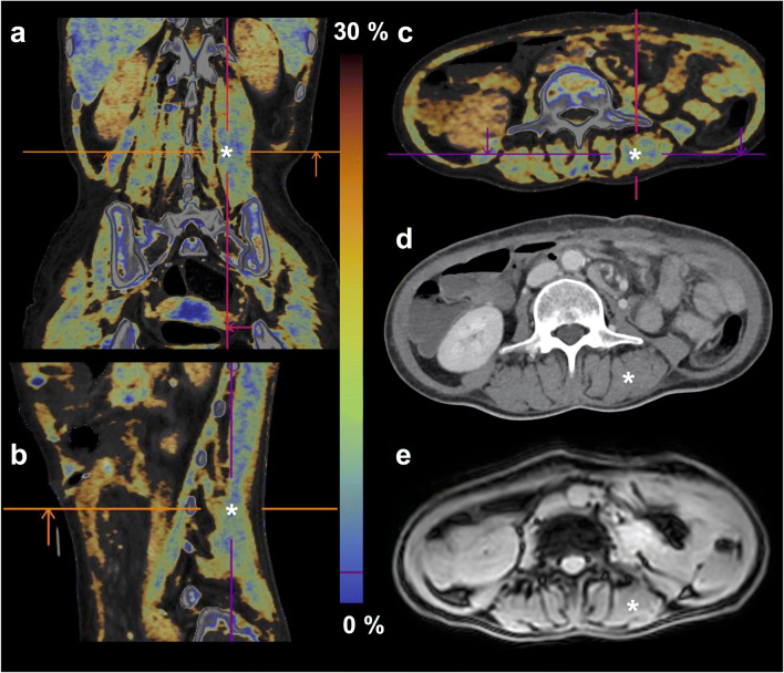
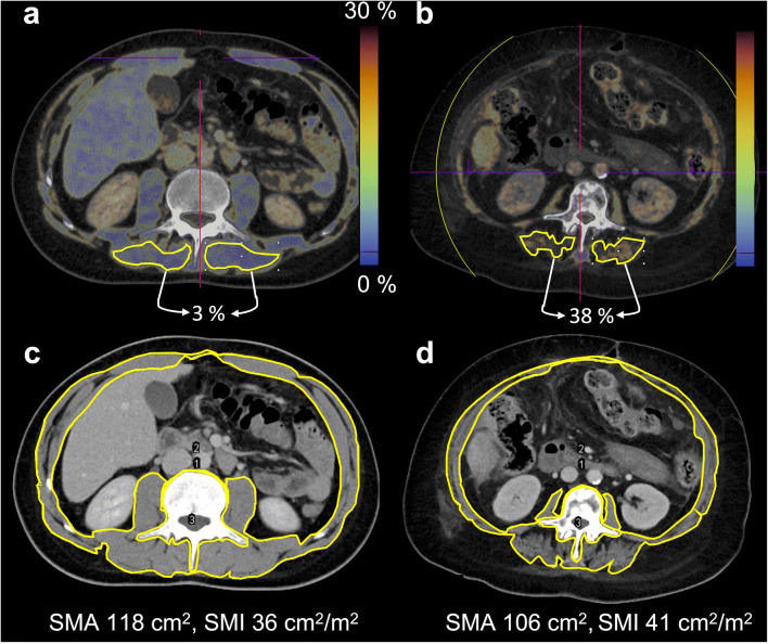
Twenty-one patients with abdominal contrast-enhanced DECT scans (100 kV/Sn 150 kV) underwent abdominal 3-T MRI. The fat fraction (DECT-FF), determined by material decomposition, and HU values on virtual non-contrast-enhanced (VNC) DECT images were measured in 126 regions of interest (≥ 6 cm) within the posterior paraspinal muscle. For validation, the MR-based fat fraction (MR-FF) was assessed by chemical shift relaxometry. Patients were categorized into groups of high or low skeletal muscle mean radiation attenuation (SMRA) and classified as either sarcopenic or non-sarcopenic, according to the skeletal muscle index (SMI) and cut-off values from non-contrast-enhanced single-energy CT. Spearman's and intraclass correlation, Bland-Altman analysis, and mixed linear models were employed.

RESULTS

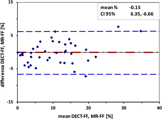
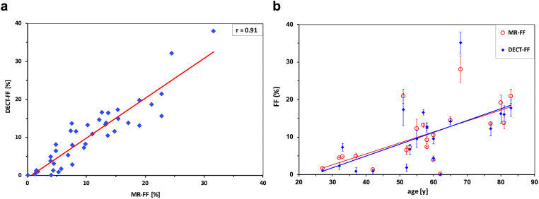
The correlation was excellent between DECT-FF and MR-FF (r = 0.91), DECT VNC HU and MR-FF (r = - 0.90), and DECT-FF and DECT VNC HU (r = - 0.98). Intraclass correlation between DECT-FF and MR-FF was good (r = 0.83 [95% CI 0.71-0.90]), with a mean difference of - 0.15% (SD 3.32 [95% CI 6.35 to - 6.66]). Categorization using the SMRA yielded an eightfold difference in DECT VNC HU values between both groups (5 HU [95% CI 23-11], 42 HU [95% CI 33-56], p = 0.05). No significant relationship between DECT-FF and SMI-based classifications was observed.

CONCLUSIONS

Fat quantification within the skeletal muscle using DECT is both feasible and reliable. DECT muscle analysis offers a new approach to determine muscle quality, which is important for the diagnosis and therapeutic monitoring of sarcopenia, as a comorbidity associated with poor clinical outcome.

KEY POINTS

• Dual-energy CT (DECT) material decomposition and virtual non-contrast-enhanced DECT HU values assess muscle fat reliably. • Virtual non-contrast-enhanced dual-energy CT HU values allow to differentiate between high and low native skeletal muscle mean radiation attenuation in contrast-enhanced DECT scans. • Measuring muscle fat by dual-energy computed tomography is a new approach for the determination of muscle quality, an important parameter for the diagnostic confirmation of sarcopenia as a comorbidity associated with poor clinical outcome.

摘要

目的

使用双能 CT(DECT)定量测量骨骼肌内的脂肪比例,作为肌肉质量的指标,并通过 MRI 对该方法进行验证。

方法

21 名患者行腹部增强 DECT 扫描(100 kV/Sn 150 kV),同时进行腹部 3-T MRI 检查。通过物质分解法测量 126 个感兴趣区(≥6cm)的后脊柱旁肌内的脂肪分数(DECT-FF),并在虚拟非增强(VNC)DECT 图像上测量 CT 值(HU 值)。为了验证,通过化学位移弛豫法评估基于 MRI 的脂肪分数(MR-FF)。根据骨骼肌指数(SMI)和非增强单能 CT 的截断值,将患者分为高或低骨骼肌平均辐射衰减(SMRA)组,并分类为肌少症或非肌少症。采用 Spearman 相关分析、组内相关分析、Bland-Altman 分析和混合线性模型。

结果

DECT-FF 与 MR-FF(r = 0.91)、DECT VNC HU 值与 MR-FF(r = -0.90)、DECT-FF 与 DECT VNC HU 值(r = -0.98)之间的相关性均极好。DECT-FF 与 MR-FF 之间的组内相关度较好(r = 0.83[95%CI 0.71-0.90]),平均差值为 -0.15%(SD 3.32[95%CI 6.35-6.66])。使用 SMRA 进行分类,两组之间的 DECT VNC HU 值差异高达 8 倍(5 HU[95%CI 23-11],42 HU[95%CI 33-56],p = 0.05)。未观察到 DECT-FF 与基于 SMI 的分类之间存在显著关系。

结论

使用 DECT 定量测量骨骼肌内的脂肪是可行且可靠的。DECT 肌肉分析为确定肌肉质量提供了一种新方法,这对于肌少症的诊断和治疗监测很重要,因为肌少症是一种与不良临床结局相关的合并症。

关键点

  1. 双能 CT(DECT)物质分解和虚拟非增强 DECT HU 值可可靠地评估肌肉脂肪。

  2. 虚拟非增强双能 CT HU 值可区分增强 DECT 扫描中高和低的固有骨骼肌平均辐射衰减。

  3. 通过双能计算机断层扫描测量肌肉脂肪是确定肌肉质量的一种新方法,肌肉质量是作为与不良临床结局相关的合并症的肌少症诊断的重要参数。

https://cdn.ncbi.nlm.nih.gov/pmc/blobs/b588/8452571/8d75e5212a59/330_2021_7820_Fig4_HTML.jpg
https://cdn.ncbi.nlm.nih.gov/pmc/blobs/b588/8452571/b840763b9ae6/330_2021_7820_Fig1_HTML.jpg
https://cdn.ncbi.nlm.nih.gov/pmc/blobs/b588/8452571/7f894d7ef86d/330_2021_7820_Fig2_HTML.jpg
https://cdn.ncbi.nlm.nih.gov/pmc/blobs/b588/8452571/deda0e97b149/330_2021_7820_Fig3_HTML.jpg
https://cdn.ncbi.nlm.nih.gov/pmc/blobs/b588/8452571/8d75e5212a59/330_2021_7820_Fig4_HTML.jpg

相似文献

1
Skeletal muscle fat quantification by dual-energy computed tomography in comparison with 3T MR imaging.双能 CT 与 3T MRI 对骨骼肌脂肪定量的比较研究。
Eur Radiol. 2021 Oct;31(10):7529-7539. doi: 10.1007/s00330-021-07820-1. Epub 2021 Mar 26.
2
Dual-Energy CT muscle fat fraction as a new imaging biomarker of body composition and survival predictor in critically ill patients.双能 CT 肌肉脂肪分数作为一种新的成像生物标志物,用于评估危重症患者的身体成分和预测生存情况。
Eur Radiol. 2024 Nov;34(11):7408-7418. doi: 10.1007/s00330-024-10779-4. Epub 2024 May 22.
3
Fat Quantification in Dual-Layer Detector Spectral Computed Tomography: Experimental Development and First In-Patient Validation.双层探测器光谱 CT 中的脂肪定量:实验开发与首次临床验证。
Invest Radiol. 2022 Jul 1;57(7):463-469. doi: 10.1097/RLI.0000000000000858. Epub 2022 Feb 11.
4
Tin-filter enhanced dual-energy-CT: image quality and accuracy of CT numbers in virtual noncontrast imaging.锡滤器增强双能量 CT:虚拟非对比成像中 CT 数的图像质量和准确性。
Acad Radiol. 2013 May;20(5):596-603. doi: 10.1016/j.acra.2013.01.010. Epub 2013 Mar 13.
5
Quantitative attenuation accuracy of virtual non-enhanced imaging compared to that of true non-enhanced imaging on dual-source dual-energy CT.双源双能 CT 虚拟平扫与真实平扫的定量衰减准确性比较。
Abdom Radiol (NY). 2020 Apr;45(4):1100-1109. doi: 10.1007/s00261-020-02415-8.
6
Virtual non-contrast images calculated from dual-energy CT shoulder arthrography improve the detection of intraarticular loose bodies.基于双能 CT 肩关节造影的虚拟非对比图像可提高关节内游离体的检出率。
Skeletal Radiol. 2022 Aug;51(8):1639-1647. doi: 10.1007/s00256-022-04007-7. Epub 2022 Feb 11.
7
Attenuation values of renal parenchyma in virtual noncontrast images acquired from multiphase renal dual-energy CT: Comparison with standard noncontrast CT.多期肾双能 CT 虚拟非对比图像中肾实质衰减值:与标准非对比 CT 的比较。
Eur J Radiol. 2018 Apr;101:103-110. doi: 10.1016/j.ejrad.2018.02.001. Epub 2018 Feb 7.
8
Dual Energy-Derived Metrics for Differentiating Adrenal Adenomas From Nonadenomas on Single-Phase Contrast-Enhanced CT.基于双能量 CT 衍生指标鉴别单相增强 CT 扫描中肾上腺腺瘤与非腺瘤。
AJR Am J Roentgenol. 2023 May;220(5):693-704. doi: 10.2214/AJR.22.28323. Epub 2022 Oct 23.
9
Fat quantification of the rotator cuff muscles using CT histogram analysis in comparison with Dixon MRI sequence.使用 CT 直方图分析对肩袖肌肉进行脂肪定量与 Dixon MRI 序列的比较。
Br J Radiol. 2023 Oct;96(1150):20220817. doi: 10.1259/bjr.20220817. Epub 2023 Sep 3.
10
Potential of gadolinium as contrast material in second generation dual energy computed tomography - An ex vivo phantom study.钆作为第二代双能计算机断层扫描造影剂的潜力——一项离体模体研究。
Clin Imaging. 2017 May-Jun;43:74-79. doi: 10.1016/j.clinimag.2017.02.005. Epub 2017 Feb 22.

引用本文的文献

1
Prognostic significance of preoperative abdominal fat thickness for pancreatic ductal adenocarcinoma.术前腹部脂肪厚度对胰腺导管腺癌的预后意义
Discov Oncol. 2025 Jun 20;16(1):1163. doi: 10.1007/s12672-025-02987-1.
2
Changes of water and muscle content in lumbar paraspinal muscle degeneration and gender differences during aging using dual-energy CT.使用双能CT评估衰老过程中腰椎旁肌退变时水分和肌肉含量的变化及性别差异。
Eur Spine J. 2025 Jun 5. doi: 10.1007/s00586-025-08968-w.
3
Differences in muscle morphology assessed by ultrasound at various muscle regions and their impact on voluntary and involuntary force production.

本文引用的文献

1
Preoperative sarcopenia is associated with poor overall survival in pancreatic cancer patients following pancreaticoduodenectomy.术前肌少症与胰十二指肠切除术后胰腺癌患者的总体生存不良相关。
Eur Radiol. 2021 Apr;31(4):2472-2481. doi: 10.1007/s00330-020-07294-7. Epub 2020 Sep 24.
2
Dual-Energy Computed Tomography for Fat Quantification in the Liver and Bone Marrow: A Literature Review.双能 CT 用于肝脏和骨髓脂肪定量的文献综述。
Rofo. 2020 Dec;192(12):1137-1153. doi: 10.1055/a-1212-6017. Epub 2020 Sep 10.
3
Assessing skeletal muscle radiodensity by computed tomography: An integrative review of the applied methodologies.
通过超声评估的不同肌肉区域的肌肉形态差异及其对随意和非随意力量产生的影响。
Sci Rep. 2025 Mar 18;15(1):9247. doi: 10.1038/s41598-025-94158-z.
4
Myosteatosis: diagnostic significance and assessment by imaging approaches.肌脂肪变性:影像学方法的诊断意义及评估
Quant Imaging Med Surg. 2024 Nov 1;14(11):7937-7957. doi: 10.21037/qims-24-365. Epub 2024 Oct 8.
5
What to Expect (and What Not) from Dual-Energy CT Imaging Now and in the Future?双能CT成像的现状与未来:我们能期待什么(以及不能期待什么)?
J Imaging. 2024 Jun 26;10(7):154. doi: 10.3390/jimaging10070154.
6
Role of effective atomic number of paraspinal muscles in the prediction of acute vertebral fracture risk assessment: a cross-sectional case-control study.椎旁肌肉有效原子序数在急性椎体骨折风险评估预测中的作用:一项横断面病例对照研究。
Br J Radiol. 2024 Aug 1;97(1160):1437-1442. doi: 10.1093/bjr/tqae112.
7
Fat quantification in dual-layer detector spectral CT: How to handle iron overload, varying tube voltage and radiation dose Indices.双层探测器光谱 CT 中的脂肪定量:如何处理铁过载、管电压变化和辐射剂量指数。
PLoS One. 2024 May 23;19(5):e0302863. doi: 10.1371/journal.pone.0302863. eCollection 2024.
8
Dual-Energy CT muscle fat fraction as a new imaging biomarker of body composition and survival predictor in critically ill patients.双能 CT 肌肉脂肪分数作为一种新的成像生物标志物,用于评估危重症患者的身体成分和预测生存情况。
Eur Radiol. 2024 Nov;34(11):7408-7418. doi: 10.1007/s00330-024-10779-4. Epub 2024 May 22.
9
SAROS: A dataset for whole-body region and organ segmentation in CT imaging.SAROS:用于 CT 成像中全身区域和器官分割的数据集。
Sci Data. 2024 May 10;11(1):483. doi: 10.1038/s41597-024-03337-6.
10
Dual-energy CT applications in musculoskeletal disorders.双能 CT 在肌肉骨骼疾病中的应用。
Br J Radiol. 2024 Mar 28;97(1156):705-715. doi: 10.1093/bjr/tqae023.
通过计算机断层扫描评估骨骼肌放射性密度:应用方法的综合综述。
Clin Physiol Funct Imaging. 2020 Jul;40(4):207-223. doi: 10.1111/cpf.12629. Epub 2020 Apr 14.
4
Impact of sarcopenia in trauma and surgical patient population: A literature review.创伤和外科患者人群中肌少症的影响:文献综述。
Asian J Surg. 2020 Jun;43(6):647-653. doi: 10.1016/j.asjsur.2019.10.010. Epub 2019 Nov 30.
5
CT-assessed sarcopenia is a predictive factor for both long-term and short-term outcomes in gastrointestinal oncology patients: a systematic review and meta-analysis.CT 评估的肌肉减少症是胃肠道肿瘤患者长期和短期结局的预测因素:系统评价和荟萃分析。
Cancer Imaging. 2019 Dec 3;19(1):82. doi: 10.1186/s40644-019-0270-0.
6
Single-slice CT measurements allow for accurate assessment of sarcopenia and body composition.单层面 CT 测量可准确评估肌肉减少症和身体成分。
Eur Radiol. 2020 Mar;30(3):1701-1708. doi: 10.1007/s00330-019-06526-9. Epub 2019 Nov 27.
7
Cachexia and Sarcopenia in Older Adults with Cancer: A Comprehensive Review.老年癌症患者的恶病质和肌肉减少症:一项综合综述。
Cancers (Basel). 2019 Nov 25;11(12):1861. doi: 10.3390/cancers11121861.
8
Association of sarcopenic obesity with the risk of all-cause mortality among adults over a broad range of different settings: a updated meta-analysis.在广泛的不同环境中,与成年人全因死亡率相关的肌少症性肥胖的关联:一项更新的荟萃分析。
BMC Geriatr. 2019 Jul 3;19(1):183. doi: 10.1186/s12877-019-1195-y.
9
Sarcopenia.肌肉减少症。
Lancet. 2019 Jun 29;393(10191):2636-2646. doi: 10.1016/S0140-6736(19)31138-9. Epub 2019 Jun 3.
10
Virtual unenhanced phase with spectral dual-energy CT: Is it an alternative to conventional true unenhanced phase for abdominal tissues?虚拟平扫期双能 CT 光谱成像:能否替代腹部组织常规的真实平扫期?
Diagn Interv Imaging. 2019 Sep;100(9):503-511. doi: 10.1016/j.diii.2019.04.007. Epub 2019 May 31.