Medicine, Hematology-Oncology, David Geffen School of Medicine, University of California Los Angeles, Los Angeles, California.
Computational Medicine, David Geffen School of Medicine, University of California Los Angeles, Los Angeles, California.
Cancer Epidemiol Biomarkers Prev. 2021 Jun;30(6):1241-1249. doi: 10.1158/1055-9965.EPI-20-1297. Epub 2021 Mar 26.
Estrogens are thought to contribute to breast cancer risk through cell cycling and accelerated breast aging. We hypothesize that lifetime estrogen exposure drives early epigenetic breast aging observed in healthy women. In this study, we examined associations between hormonal factors and epigenetic aging measures in healthy breast tissues.
We extracted DNA from breast tissue specimens from 192 healthy female donors to the Susan G. Komen Tissue Bank at the Indiana University Simon Cancer Center. Methylation experiments were performed using the Illumina EPIC 850K array platform. Age-adjusted regression models were used to examine for associations between factors related to estrogen exposure and five DNA methylation-based estimates: Grim age, pan-tissue age, Hannum age, phenotypic age, and skin and blood clock age.
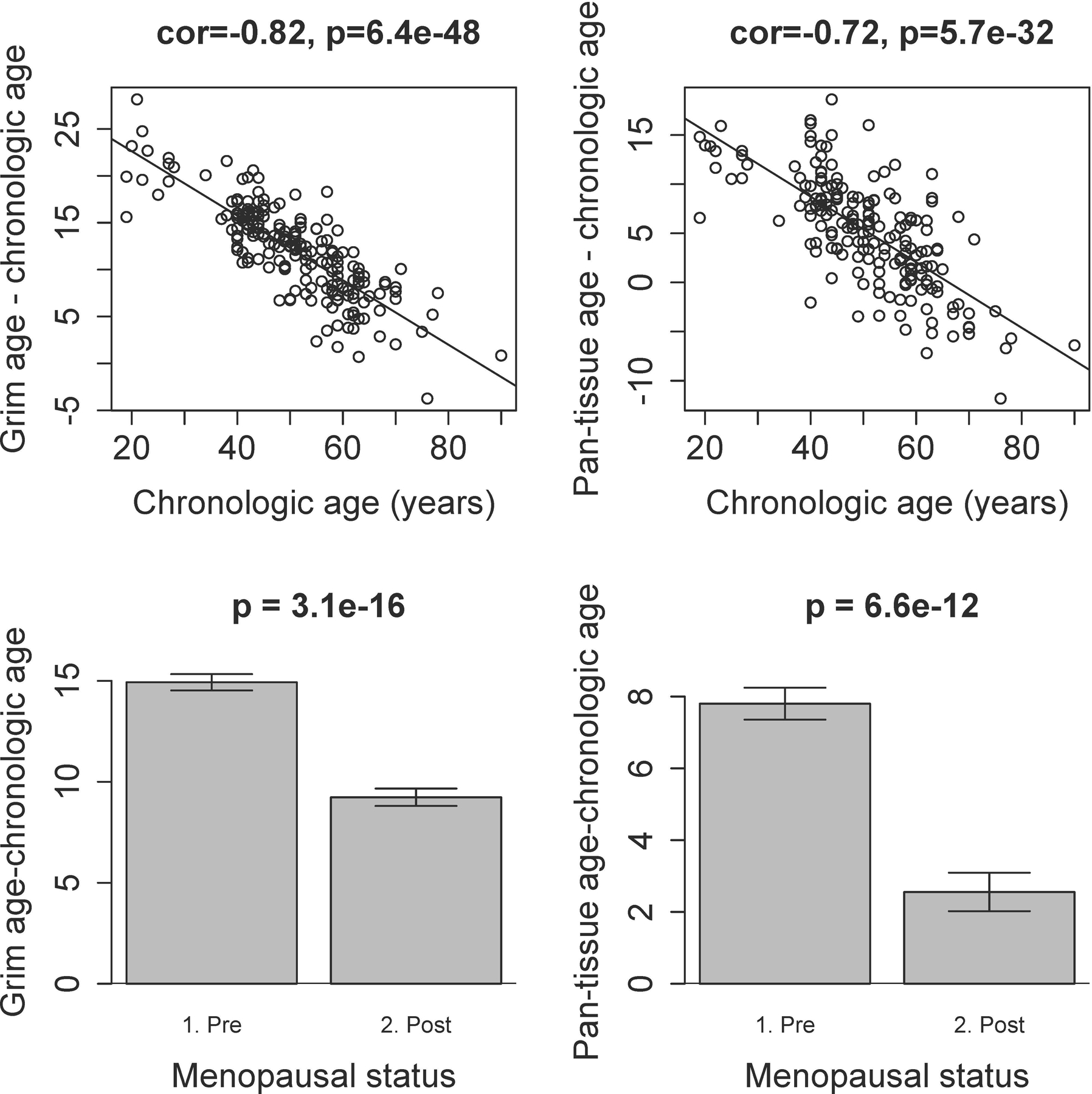
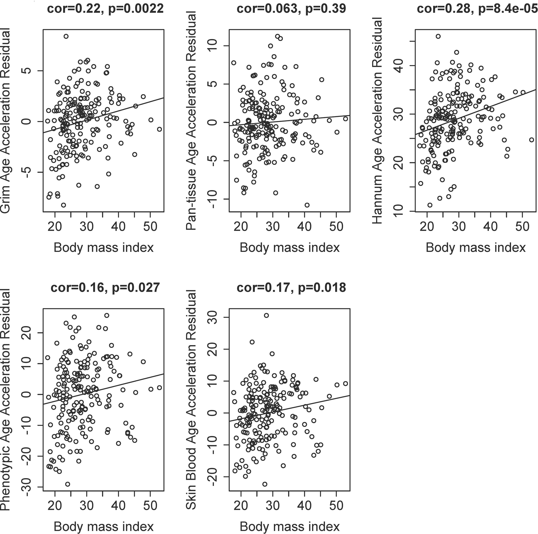
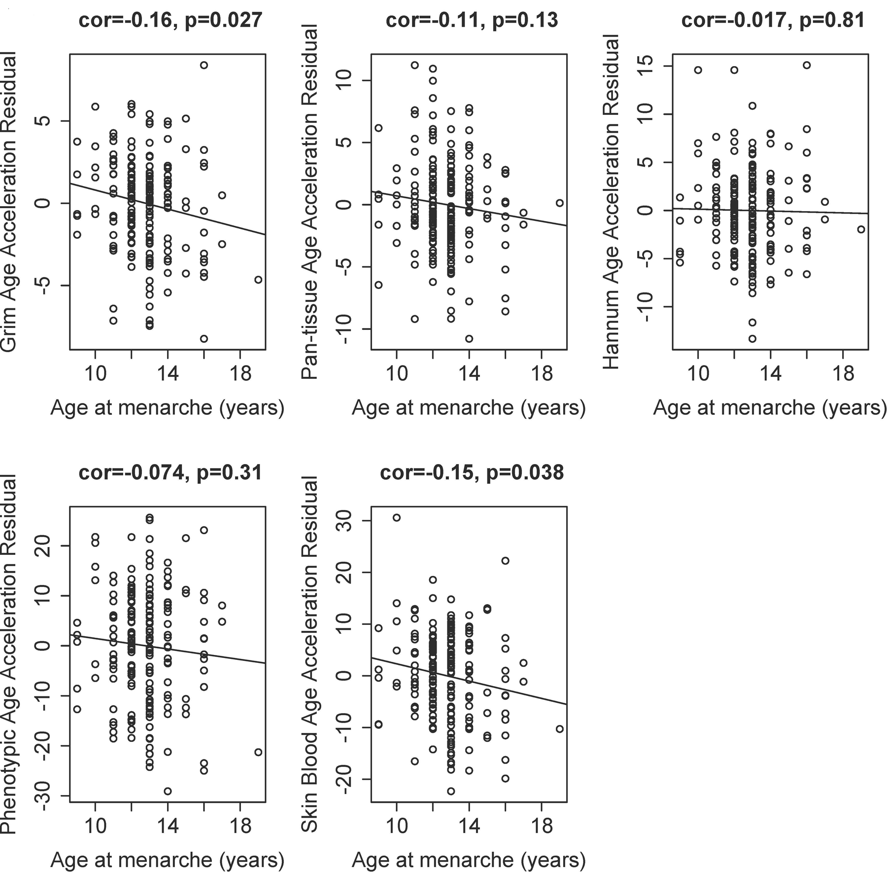
Women were aged 19-90 years, with 95 premenopausal, and 97 nulliparous women. The age difference (Grim age - chronologic age) was higher at earlier ages close to menarche. We found significant associations between earlier age at menarche and age-adjusted accelerations according to the Grim clock, the skin and blood clock, and between higher body mass index (BMI) and age-adjusted accelerations in the Grim clock, Hannum clock, phenotypic clock, and skin and blood clock.
Earlier age at menarche and higher BMI are associated with elevations in DNA methylation-based age estimates in healthy breast tissues, suggesting that cumulative estrogen exposure drives breast epigenetic aging.
Epigenetic clock measures may help advance inquiry into the relationship between accelerated breast tissue aging and an elevated incidence of breast cancer in younger women.
雌激素被认为通过细胞周期和加速乳腺衰老来增加乳腺癌风险。我们假设终生雌激素暴露会导致健康女性中观察到的早期表观遗传乳腺衰老。在这项研究中,我们研究了激素因素与健康乳腺组织中表观遗传衰老指标之间的关联。
我们从印第安纳大学西蒙癌症中心苏珊·科曼组织库的 192 名健康女性供体中提取了乳腺组织标本的 DNA。使用 Illumina EPIC 850K 芯片平台进行了甲基化实验。使用年龄调整的回归模型,研究了与雌激素暴露相关的因素与五种基于 DNA 甲基化的估计值(Grim 年龄、泛组织年龄、Hannum 年龄、表型年龄和皮肤及血液时钟年龄)之间的关联。
女性年龄为 19-90 岁,其中 95 名处于绝经前,97 名未生育。在接近初潮的早期,年龄差异(Grim 年龄-实际年龄)较大。我们发现,初潮年龄较早与 Grim 时钟、皮肤和血液时钟的年龄调整加速以及体重指数(BMI)较高与 Grim 时钟、Hannum 时钟、表型时钟和皮肤及血液时钟的年龄调整加速之间存在显著关联。
初潮年龄较早和 BMI 较高与健康乳腺组织中基于 DNA 甲基化的年龄估计值升高有关,提示累积雌激素暴露会导致乳腺表观遗传衰老。
表观遗传时钟测量值可能有助于深入研究加速的乳腺组织衰老与年轻女性中乳腺癌发病率升高之间的关系。