• 文献检索
  • 文档翻译
  • 深度研究
  • 学术资讯
  • Suppr Zotero 插件Zotero 插件
  • 邀请有礼
  • 套餐&价格
  • 历史记录
应用&插件
Suppr Zotero 插件Zotero 插件浏览器插件Mac 客户端Windows 客户端微信小程序
定价
高级版会员购买积分包购买API积分包
服务
文献检索文档翻译深度研究API 文档MCP 服务
关于我们
关于 Suppr公司介绍联系我们用户协议隐私条款
关注我们

Suppr 超能文献

核心技术专利:CN118964589B侵权必究
粤ICP备2023148730 号-1Suppr @ 2026

文献检索

告别复杂PubMed语法,用中文像聊天一样搜索,搜遍4000万医学文献。AI智能推荐,让科研检索更轻松。

立即免费搜索

文件翻译

保留排版,准确专业,支持PDF/Word/PPT等文件格式,支持 12+语言互译。

免费翻译文档

深度研究

AI帮你快速写综述,25分钟生成高质量综述,智能提取关键信息,辅助科研写作。

立即免费体验

用于大规模免疫反应成像的学习自适应多光子照明显微镜。

Learned adaptive multiphoton illumination microscopy for large-scale immune response imaging.

作者信息

Pinkard Henry, Baghdassarian Hratch, Mujal Adriana, Roberts Ed, Hu Kenneth H, Friedman Daniel Haim, Malenica Ivana, Shagam Taylor, Fries Adam, Corbin Kaitlin, Krummel Matthew F, Waller Laura

机构信息

Department of Electrical Engineering and Computer Sciences, University of California, Berkeley, CA, USA.

Computational Biology Graduate Group, University of California, Berkeley, CA, USA.

出版信息

Nat Commun. 2021 Mar 26;12(1):1916. doi: 10.1038/s41467-021-22246-5.

DOI:10.1038/s41467-021-22246-5
PMID:33772022
原文链接:https://pmc.ncbi.nlm.nih.gov/articles/PMC7997974/
Abstract

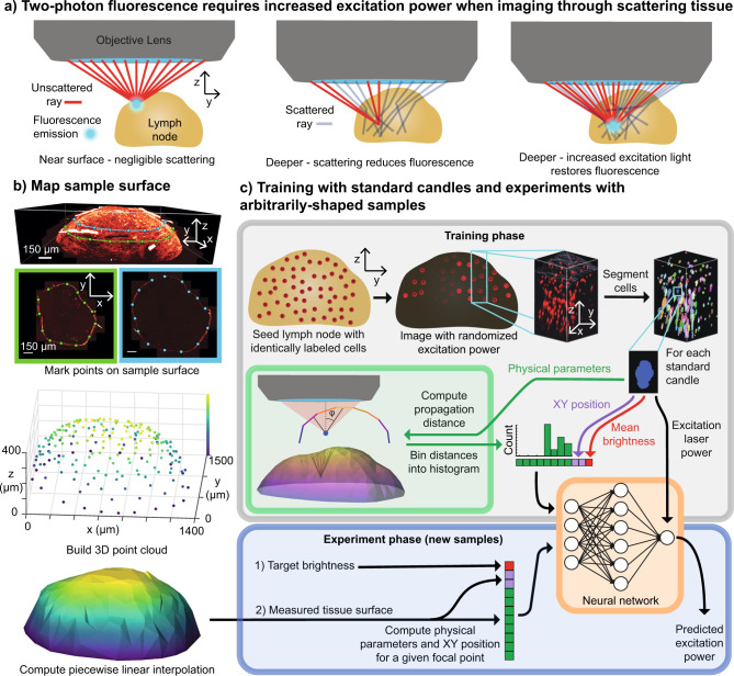
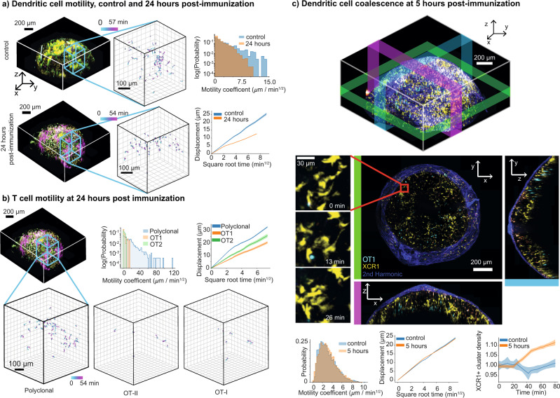
Multiphoton microscopy is a powerful technique for deep in vivo imaging in scattering samples. However, it requires precise, sample-dependent increases in excitation power with depth in order to generate contrast in scattering tissue, while minimizing photobleaching and phototoxicity. We show here how adaptive imaging can optimize illumination power at each point in a 3D volume as a function of the sample's shape, without the need for specialized fluorescent labeling. Our method relies on training a physics-based machine learning model using cells with identical fluorescent labels imaged in situ. We use this technique for in vivo imaging of immune responses in mouse lymph nodes following vaccination. We achieve visualization of physiologically realistic numbers of antigen-specific T cells (~2 orders of magnitude lower than previous studies), and demonstrate changes in the global organization and motility of dendritic cell networks during the early stages of the immune response. We provide a step-by-step tutorial for implementing this technique using exclusively open-source hardware and software.

摘要

多光子显微镜是一种用于散射样本体内深度成像的强大技术。然而,为了在散射组织中产生对比度,同时将光漂白和光毒性降至最低,它需要根据样本情况精确地随着深度增加激发功率。我们在此展示了自适应成像如何根据样本形状在三维体积中的每个点优化照明功率,而无需专门的荧光标记。我们的方法依赖于使用原位成像的具有相同荧光标记的细胞来训练基于物理的机器学习模型。我们将此技术用于疫苗接种后小鼠淋巴结免疫反应的体内成像。我们实现了对抗抗原特异性T细胞生理现实数量的可视化(比以前的研究低约2个数量级),并证明了免疫反应早期树突状细胞网络的整体组织和运动性的变化。我们提供了一个使用完全开源硬件和软件实施此技术的分步教程。

https://cdn.ncbi.nlm.nih.gov/pmc/blobs/c0c6/7997974/943a8b3e9e91/41467_2021_22246_Fig3_HTML.jpg
https://cdn.ncbi.nlm.nih.gov/pmc/blobs/c0c6/7997974/46a8721063b5/41467_2021_22246_Fig1_HTML.jpg
https://cdn.ncbi.nlm.nih.gov/pmc/blobs/c0c6/7997974/5577e233cff8/41467_2021_22246_Fig2_HTML.jpg
https://cdn.ncbi.nlm.nih.gov/pmc/blobs/c0c6/7997974/943a8b3e9e91/41467_2021_22246_Fig3_HTML.jpg
https://cdn.ncbi.nlm.nih.gov/pmc/blobs/c0c6/7997974/46a8721063b5/41467_2021_22246_Fig1_HTML.jpg
https://cdn.ncbi.nlm.nih.gov/pmc/blobs/c0c6/7997974/5577e233cff8/41467_2021_22246_Fig2_HTML.jpg
https://cdn.ncbi.nlm.nih.gov/pmc/blobs/c0c6/7997974/943a8b3e9e91/41467_2021_22246_Fig3_HTML.jpg

相似文献

1
Learned adaptive multiphoton illumination microscopy for large-scale immune response imaging.用于大规模免疫反应成像的学习自适应多光子照明显微镜。
Nat Commun. 2021 Mar 26;12(1):1916. doi: 10.1038/s41467-021-22246-5.
2
Multiphoton Intravital Microscopy of Mandibular Draining Lymph Nodes: A Mouse Model to Study Corneal Immune Responses.下颌引流淋巴结的多光子活体显微镜检查:一种研究角膜免疫反应的小鼠模型。
Front Immunol. 2020 Feb 21;11:39. doi: 10.3389/fimmu.2020.00039. eCollection 2020.
3
Multiphoton intravital microscopy to study lymphocyte motility in lymph nodes.利用多光子活体显微镜研究淋巴结中淋巴细胞的运动性。
Methods Mol Biol. 2012;757:247-57. doi: 10.1007/978-1-61779-166-6_16.
4
T cell migration dynamics within lymph nodes during steady state: an overview of extracellular and intracellular factors influencing the basal intranodal T cell motility.稳态下淋巴结内T细胞迁移动力学:影响基础淋巴结内T细胞运动性的细胞外和细胞内因素概述
Curr Top Microbiol Immunol. 2009;334:71-105. doi: 10.1007/978-3-540-93864-4_4.
5
Intravital imaging of the mouse popliteal lymph node.小鼠腘淋巴结的活体成像。
J Vis Exp. 2012 Feb 8(60):3720. doi: 10.3791/3720.
6
Infrared multiphoton microscopy: subcellular-resolved deep tissue imaging.红外多光子显微镜:亚细胞分辨深度组织成像。
Curr Opin Biotechnol. 2009 Feb;20(1):54-62. doi: 10.1016/j.copbio.2009.02.008. Epub 2009 Mar 25.
7
Live Imaging of the Skin Immune Responses: Visualization of the Contact Hypersensitivity Response.皮肤免疫反应的活体成像:接触性超敏反应的可视化
Methods Mol Biol. 2018;1763:75-85. doi: 10.1007/978-1-4939-7762-8_8.
8
In Vivo Tracking of Particulate Antigen Localization and Recognition by B Lymphocytes at Lymph Nodes.体内追踪颗粒性抗原在淋巴结中的定位以及B淋巴细胞对其的识别
Methods Mol Biol. 2018;1707:163-169. doi: 10.1007/978-1-4939-7474-0_11.
9
Visualizing the Dynamics of T Cell-Dendritic Cell Interactions in Intact Lymph Nodes by Multiphoton Confocal Microscopy.利用多光子共聚焦显微镜观察完整淋巴结中 T 细胞与树突状细胞相互作用的动态过程。
Methods Mol Biol. 2021;2304:243-263. doi: 10.1007/978-1-0716-1402-0_13.
10
Light sheet fluorescence microscopy for in situ cell interaction analysis in mouse lymph nodes.用于小鼠淋巴结原位细胞相互作用分析的光片荧光显微镜技术
J Immunol Methods. 2016 Apr;431:1-10. doi: 10.1016/j.jim.2016.01.015. Epub 2016 Feb 2.

引用本文的文献

1
Live-cell imaging powered by computation.基于计算的活细胞成像。
Nat Rev Mol Cell Biol. 2024 Jun;25(6):443-463. doi: 10.1038/s41580-024-00702-6. Epub 2024 Feb 20.
2
Resource allocation in mammalian systems.哺乳动物系统中的资源分配。
Biotechnol Adv. 2024 Mar-Apr;71:108305. doi: 10.1016/j.biotechadv.2023.108305. Epub 2024 Jan 11.
3
Adaptive Design of Fluorescence Imaging Systems for Custom Resolution, Fields of View, and Geometries.用于定制分辨率、视场和几何形状的荧光成像系统的自适应设计。

本文引用的文献

1
De-scattering with Excitation Patterning enables rapid wide-field imaging through scattering media.采用激发图案化的去散射技术可实现通过散射介质的快速宽视场成像。
Sci Adv. 2021 Jul 7;7(28). doi: 10.1126/sciadv.aay5496. Print 2021 Jul.
2
SciPy 1.0: fundamental algorithms for scientific computing in Python.SciPy 1.0:Python 中的科学计算基础算法。
Nat Methods. 2020 Mar;17(3):261-272. doi: 10.1038/s41592-019-0686-2. Epub 2020 Feb 3.
3
An adaptive excitation source for high-speed multiphoton microscopy.高速多光子显微镜的自适应激励源。
BME Front. 2023 Jan 13;4:0005. doi: 10.34133/bmef.0005. eCollection 2023.
4
Light-sheets and smart microscopy, an exciting future is dawning.光片和智能显微镜,一个激动人心的未来正在到来。
Commun Biol. 2023 May 9;6(1):502. doi: 10.1038/s42003-023-04857-4.
5
An open-source microscopy framework for simultaneous control of image acquisition, reconstruction, and analysis.一个用于同时控制图像采集、重建和分析的开源显微镜框架。
HardwareX. 2023 Feb 6;13:e00400. doi: 10.1016/j.ohx.2023.e00400. eCollection 2023 Mar.
6
Event-driven acquisition for content-enriched microscopy.基于事件驱动的获取技术在富含内容的显微镜中的应用
Nat Methods. 2022 Oct;19(10):1262-1267. doi: 10.1038/s41592-022-01589-x. Epub 2022 Sep 8.
7
Event-triggered STED imaging.事件触发的 STED 成像。
Nat Methods. 2022 Oct;19(10):1268-1275. doi: 10.1038/s41592-022-01588-y. Epub 2022 Sep 8.
Nat Methods. 2020 Feb;17(2):163-166. doi: 10.1038/s41592-019-0663-9. Epub 2019 Dec 2.
4
Content-aware image restoration: pushing the limits of fluorescence microscopy.内容感知图像恢复:推动荧光显微镜的极限。
Nat Methods. 2018 Dec;15(12):1090-1097. doi: 10.1038/s41592-018-0216-7. Epub 2018 Nov 26.
5
Aberration correction considering curved sample surface shape for non-contact two-photon excitation microscopy with spatial light modulator.基于空间光调制器的非接触式双光子激发显微镜中考虑弯曲样品表面形状的像差校正。
Sci Rep. 2018 Jun 18;8(1):9252. doi: 10.1038/s41598-018-27693-7.
6
Adaptive optical fluorescence microscopy.自适应光学荧光显微镜。
Nat Methods. 2017 Mar 31;14(4):374-380. doi: 10.1038/nmeth.4218.
7
CD8 T Cells Orchestrate pDC-XCR1 Dendritic Cell Spatial and Functional Cooperativity to Optimize Priming.CD8 T细胞协调浆细胞样树突状细胞与XCR1阳性树突状细胞的空间和功能协同作用以优化启动。
Immunity. 2017 Feb 21;46(2):205-219. doi: 10.1016/j.immuni.2017.01.003. Epub 2017 Feb 9.
8
Spatiotemporal Rank Filtering Improves Image Quality Compared to Frame Averaging in 2-Photon Laser Scanning Microscopy.与双光子激光扫描显微镜中的帧平均相比,时空秩滤波可提高图像质量。
PLoS One. 2016 Mar 3;11(3):e0150430. doi: 10.1371/journal.pone.0150430. eCollection 2016.
9
The smart and gentle microscope.智能且温和的显微镜。
Nat Biotechnol. 2015 Aug;33(8):815-8. doi: 10.1038/nbt.3310.
10
Deep learning.深度学习。
Nature. 2015 May 28;521(7553):436-44. doi: 10.1038/nature14539.