She Han, Zhu Yu, Deng Haoyue, Kuang Lei, Fang He, Zhang Zisen, Duan Chenyang, Ye Jiaqing, Zhang Jie, Liu Liangming, Hu Yi, Li Tao
Department of Anesthesiology, Daping Hospital, Army Medical University, Chongqing, China.
State Key Laboratory of Trauma, Burns and Combined Injury, Second Department of Research Institute of Surgery, Daping Hospital, Army Medical University, Chongqing, China.
Front Cell Dev Biol. 2021 Mar 11;9:636327. doi: 10.3389/fcell.2021.636327. eCollection 2021.
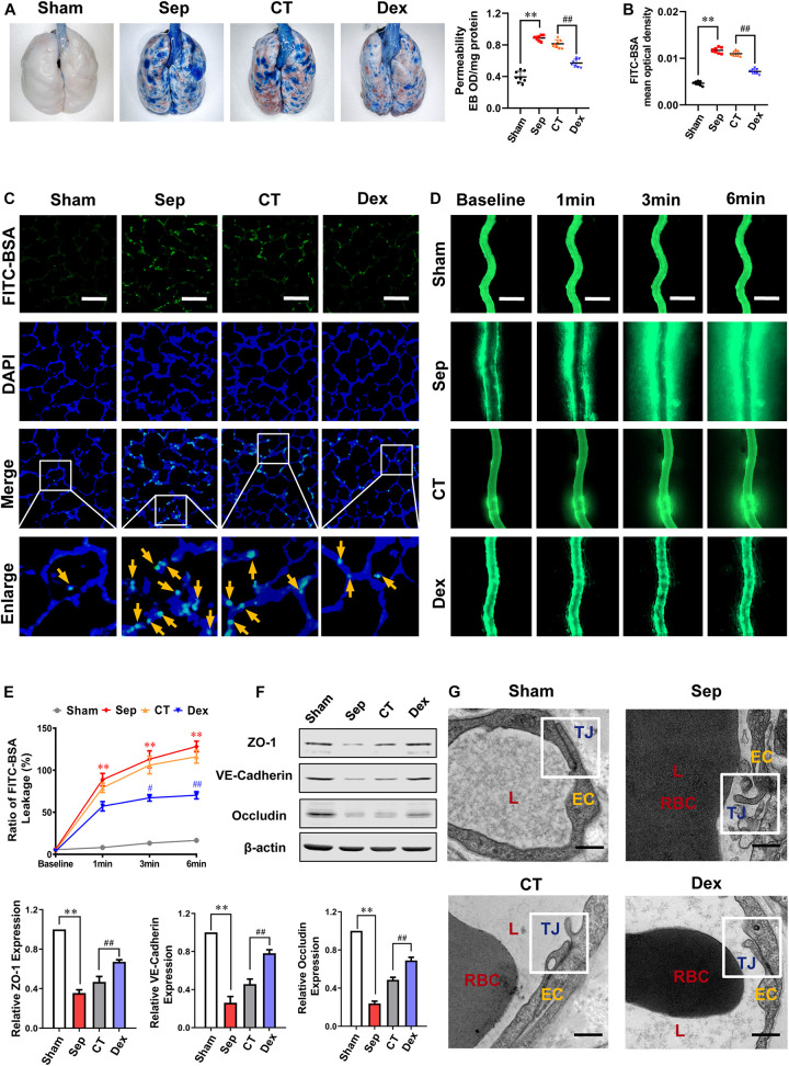
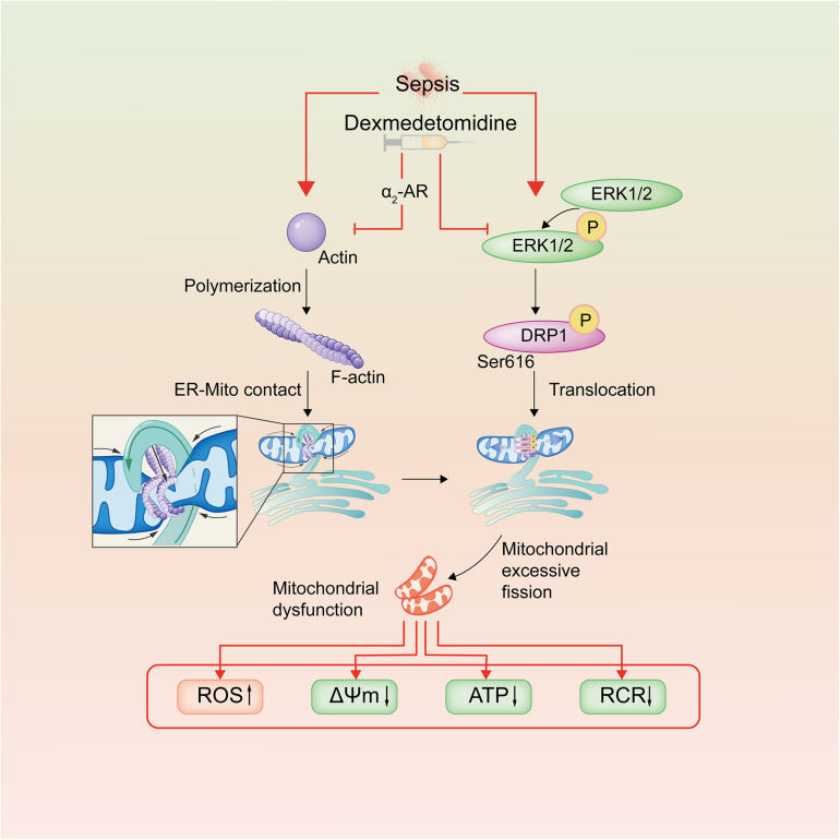
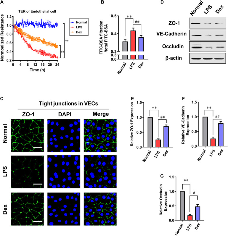
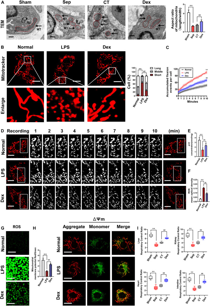
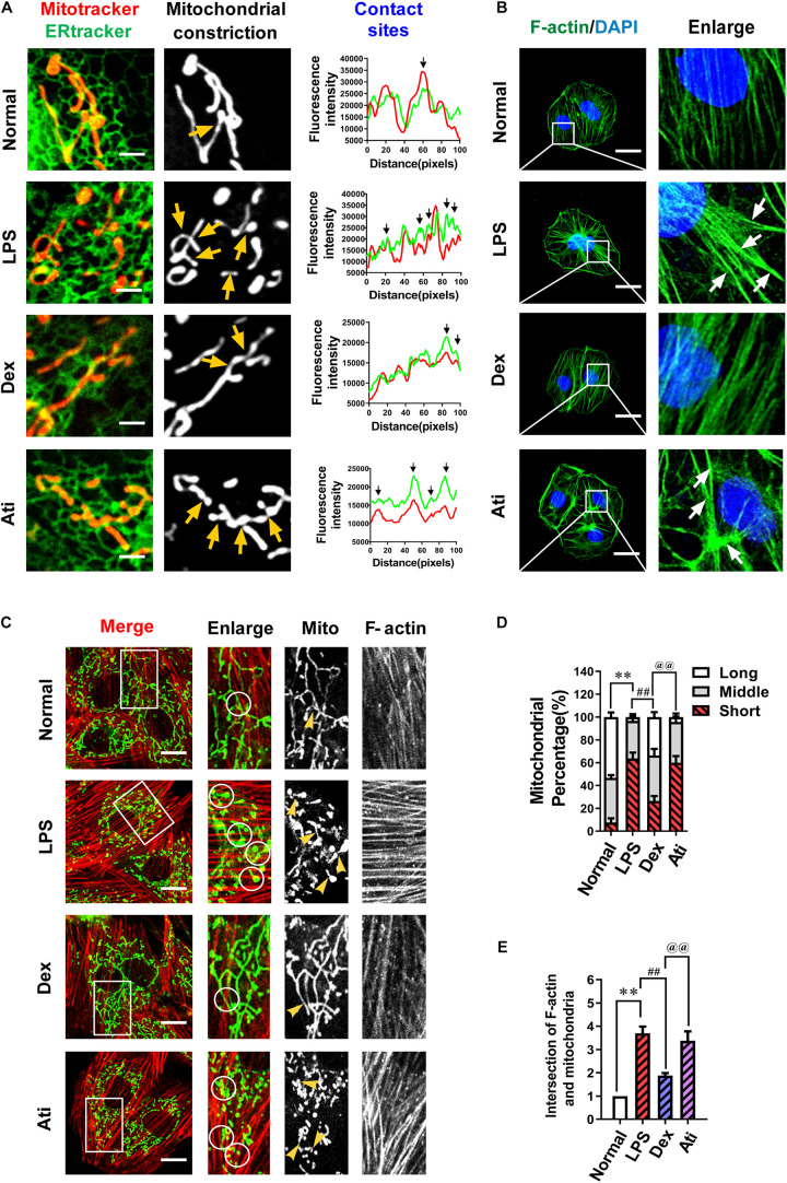
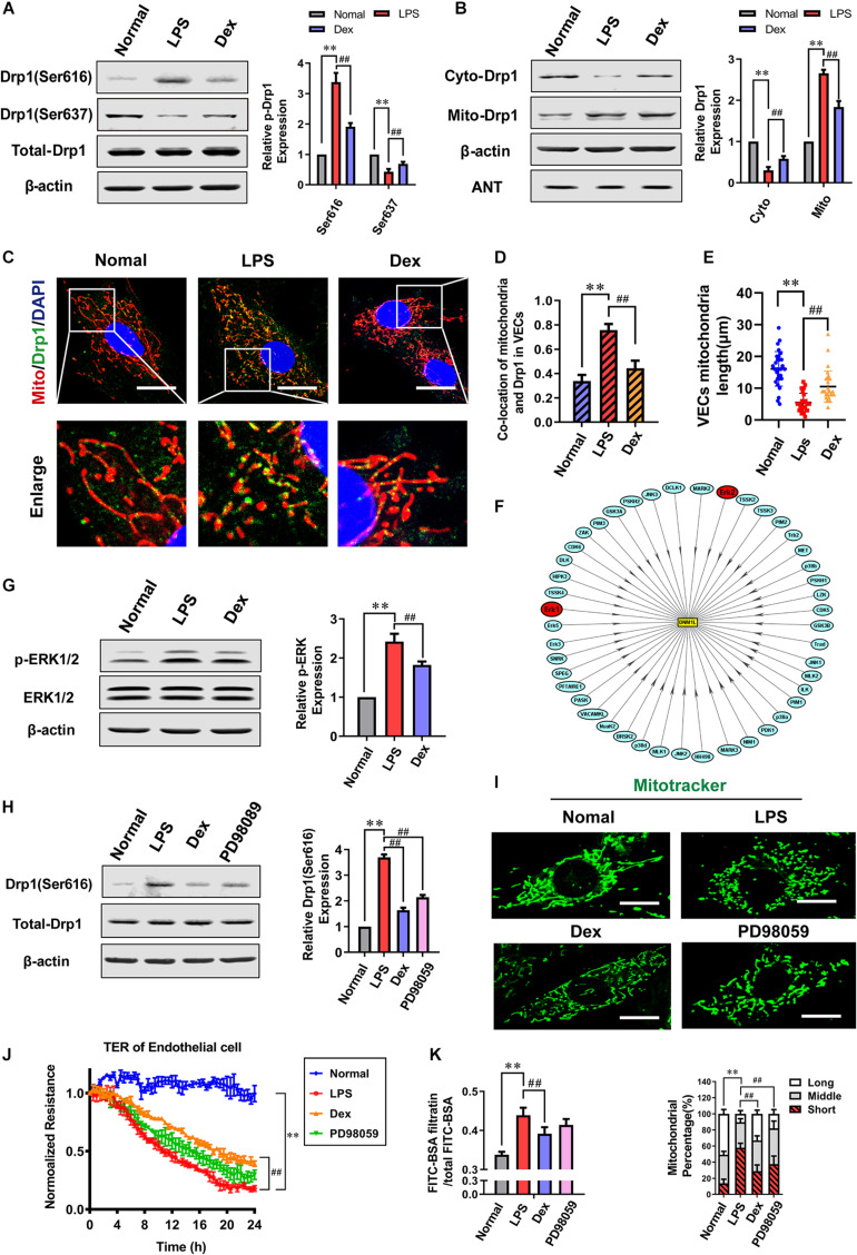
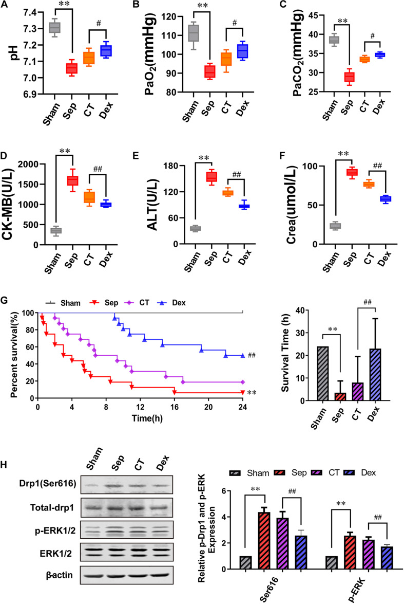
The damage of vascular endothelial barrier function induced by sepsis is critical in causing multiple organ dysfunctions. Previous studies showed that dexmedetomidine (Dex) played a vital role in protecting organ functions. However, whether Dex participates in protecting vascular leakage of sepsis and the associated underlying mechanism remains unknown yet. We used cecal ligation and puncture induced septic rats and lipopolysaccharide stimulated vascular endothelial cells (VECs) to establish models and , then the protective effects of Dex on the vascular endothelial barrier function of sepsis were observed, meanwhile, related mechanisms on regulating mitochondrial fission were further studied. The results showed that Dex could significantly reduce the permeability of pulmonary veins and mesenteric vessels, increase the expression of intercellular junction proteins, enhance the transendothelial electrical resistance and decrease the transmittance of VECs, accordingly protected organ functions and prolonged survival time in septic rats. Besides, the mitochondria of VECs were excessive division after sepsis, while Dex could significantly inhibit the mitochondrial fission and protect mitochondrial function by restoring mitochondrial morphology of VECs. Furthermore, the results showed that ER-MITO contact sites of VECs were notably increased after sepsis. Nevertheless, Dex reduced ER-MITO contact sites by regulating the polymerization of actin via α receptors. The results also found that Dex could induce the phosphorylation of the dynamin-related protein 1 through down-regulating extracellular signal-regulated kinase1/2, thus playing a role in the regulation of mitochondrial division. In conclusion, Dex has a protective effect on the vascular endothelial barrier function of septic rats. The mechanism is mainly related to the regulation of Drp1 phosphorylation of VECs, inhibition of mitochondrial division by ER-MITO contacts, and protection of mitochondrial function.
脓毒症诱导的血管内皮屏障功能损伤在导致多器官功能障碍中起关键作用。先前的研究表明,右美托咪定(Dex)在保护器官功能方面发挥着重要作用。然而,Dex是否参与保护脓毒症的血管渗漏及其相关潜在机制尚不清楚。我们使用盲肠结扎和穿刺诱导的脓毒症大鼠以及脂多糖刺激的血管内皮细胞(VECs)建立模型,然后观察Dex对脓毒症血管内皮屏障功能的保护作用,同时进一步研究调节线粒体分裂的相关机制。结果表明,Dex可显著降低肺静脉和肠系膜血管的通透性,增加细胞间连接蛋白的表达,增强跨内皮电阻并降低VECs的透光度,从而保护器官功能并延长脓毒症大鼠的存活时间。此外,脓毒症后VECs的线粒体过度分裂,而Dex可通过恢复VECs的线粒体形态显著抑制线粒体分裂并保护线粒体功能。此外,结果表明脓毒症后VECs的内质网-线粒体接触位点显著增加。然而,Dex通过α受体调节肌动蛋白的聚合来减少内质网-线粒体接触位点。结果还发现,Dex可通过下调细胞外信号调节激酶1/2诱导动力相关蛋白1的磷酸化,从而在调节线粒体分裂中发挥作用。总之,Dex对脓毒症大鼠的血管内皮屏障功能具有保护作用。其机制主要与调节VECs的Drp1磷酸化、通过内质网-线粒体接触抑制线粒体分裂以及保护线粒体功能有关。