Zhao Lidong, Zhuang Jinqiang, Wang Yihui, Zhou Dandan, Zhao Dandan, Zhu Shun, Pu Jinjun, Zhang Hongyu, Yin Ming, Zhao Wenjuan, Wang Zejian, Hong Jiang
Department of Internal and Emergency Medicine, Shanghai General Hospital, Shanghai Jiao Tong University School of Medicine (Originally Named "Shanghai First People' s Hospital"), Shanghai, China.
Department of Emergency Medicine, Putuo Hospital Affiliated to Shanghai University of Traditional Chinese Medicine, Shanghai, China.
Front Pharmacol. 2019 Feb 12;10:61. doi: 10.3389/fphar.2019.00061. eCollection 2019.
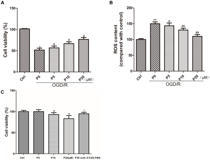
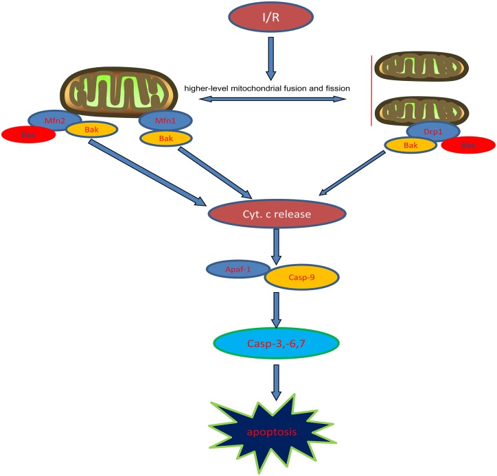
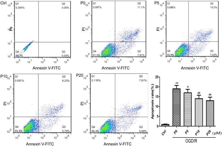
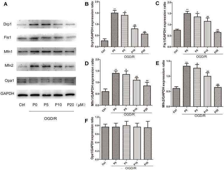
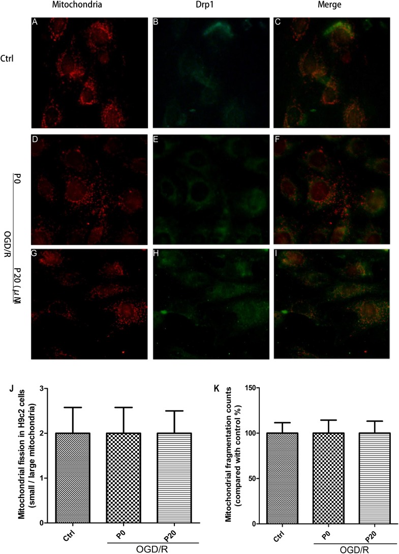
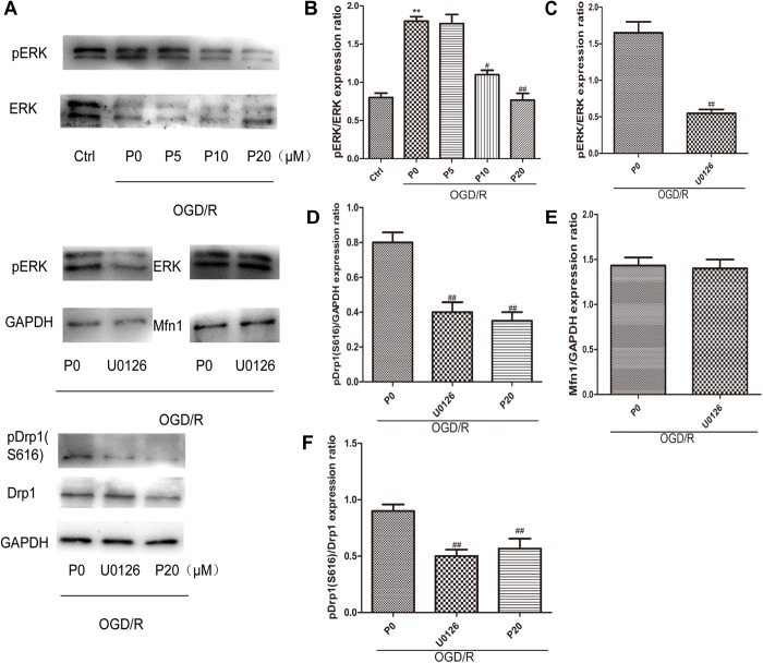
The cardioprotective effect of propofol on ischemia-reperfusion injury (I/R injury) is partly due to suppressing apoptosis. Mitochondrial dynamics are also involved in apoptosis. Mitochondrial fusion and fission lead to mitochondrial morphological changes. However, whether suppressing apoptosis effect of propofol against ischemia-reperfusion injury in the heart is via regulating mitochondrial morphology remains unclear. H9c2 cells underwent oxygen glucose deprivation (OGD) followed by reperfusion to simulate cardiomyocytes ischemia/reperfusion injury. Cell viability, apoptosis ratio and intracellular reactive oxygen species (ROS) were assessed, respectively. Mitochondrial membrane dynamin family proteins, extracellular signal regulated kinase 1 and 2 (ERK1/2), phosphorylated extracellular signal regulated kinase 1 and 2 (p-ERK1/2) and proteins related to intrinsic apoptosis pathways were detected by western blotting. The mitochondrial morphology and the distribution of dynamin-related protein 1 (Drp1) were observed by using laser confocal microscopy. Propofol enhanced the survival of H9c2 cells, decreased ROS levels and inhibited apoptosis during oxygen glucose deprivation/reperfusion (OGD/R) injury. Mitochondrial fission in H9c2 cells was inhibited by propofol during OGD injury. Propofol alleviated high levels of mitochondrial fusion and fission during OGD/R in H9c2 cells, by regulating mitochondrial membrane remodeling dynamin family proteins. Propofol inhibited Drp1 colocalization with mitochondria in H9c2 cells during OGD/R injury. Moreover, Drp1 phosphorylation was inhibited by propofol through decreasing ERK activation during OGD/R injury. We found that propofol ameliorated H9c2 cells apoptosis during OGD/R via inhibiting mitochondrial cytochrome c release and caspase-9, caspase-6, caspase-7 and caspase-3 activation. Propofol suppresses H9c2 cells apoptosis during OGD/R injury via inhibiting intrinsic apoptosis pathway, which may be partly due to reducing high levels of mitochondrial fusion and fission induced by OGD/R injury.
丙泊酚对缺血再灌注损伤(I/R损伤)的心脏保护作用部分归因于抑制细胞凋亡。线粒体动力学也参与细胞凋亡。线粒体融合和裂变导致线粒体形态变化。然而,丙泊酚对心脏缺血再灌注损伤的抑制细胞凋亡作用是否通过调节线粒体形态仍不清楚。H9c2细胞先经历氧糖剥夺(OGD),然后再灌注,以模拟心肌细胞缺血/再灌注损伤。分别评估细胞活力、凋亡率和细胞内活性氧(ROS)。通过蛋白质免疫印迹法检测线粒体膜动力蛋白家族蛋白、细胞外信号调节激酶1和2(ERK1/2)、磷酸化细胞外信号调节激酶1和2(p-ERK1/2)以及与内源性凋亡途径相关的蛋白。使用激光共聚焦显微镜观察线粒体形态和动力相关蛋白1(Drp1)的分布。丙泊酚提高了H9c2细胞在氧糖剥夺/再灌注(OGD/R)损伤期间的存活率,降低了ROS水平并抑制了细胞凋亡。在OGD损伤期间,丙泊酚抑制了H9c2细胞中的线粒体裂变。丙泊酚通过调节线粒体膜重塑动力蛋白家族蛋白,减轻了H9c2细胞在OGD/R期间高水平的线粒体融合和裂变。在OGD/R损伤期间,丙泊酚抑制了H9c2细胞中Drp1与线粒体的共定位。此外,在OGD/R损伤期间,丙泊酚通过降低ERK激活来抑制Drp1磷酸化。我们发现丙泊酚通过抑制线粒体细胞色素c释放以及半胱天冬酶-9、半胱天冬酶-6、半胱天冬酶-7和半胱天冬酶-3的激活,改善了OGD/R期间H9c2细胞的凋亡。丙泊酚通过抑制内源性凋亡途径抑制OGD/R损伤期间H9c2细胞的凋亡,这可能部分归因于减少了OGD/R损伤诱导的高水平线粒体融合和裂变。