Kauanova Sholpan, Urazbayev Arshat, Vorobjev Ivan
School of Science and Humanities, Nazarbayev University, Nur-Sultan, Kazakhstan.
National Laboratory Astana, Nazarbayev University, Nur-Sultan, Kazakhstan.
Front Cell Dev Biol. 2021 Mar 11;9:640972. doi: 10.3389/fcell.2021.640972. eCollection 2021.
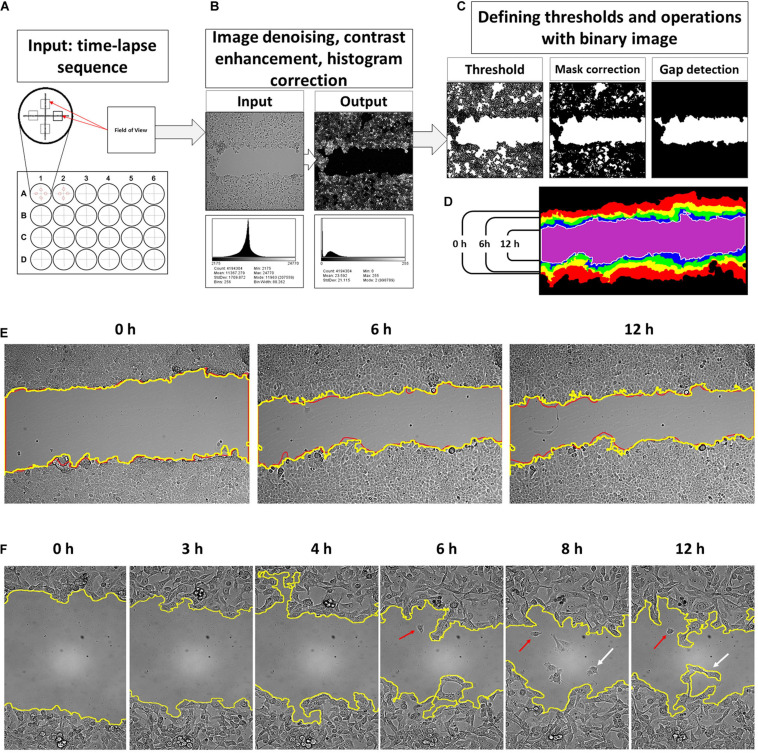
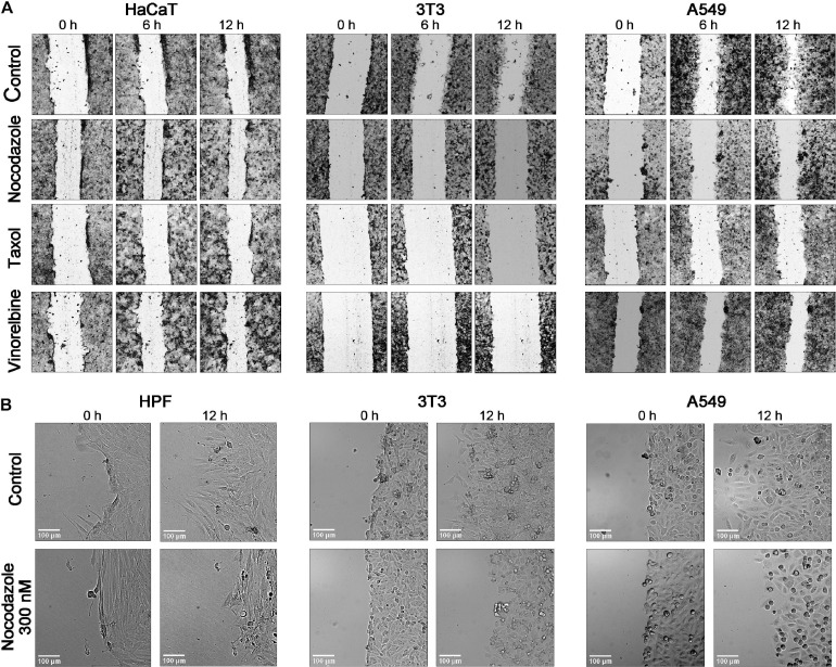
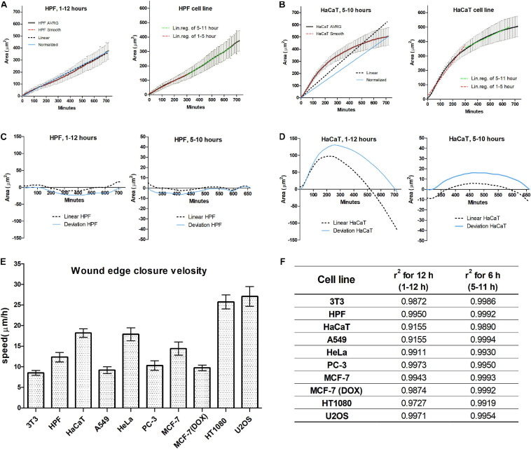
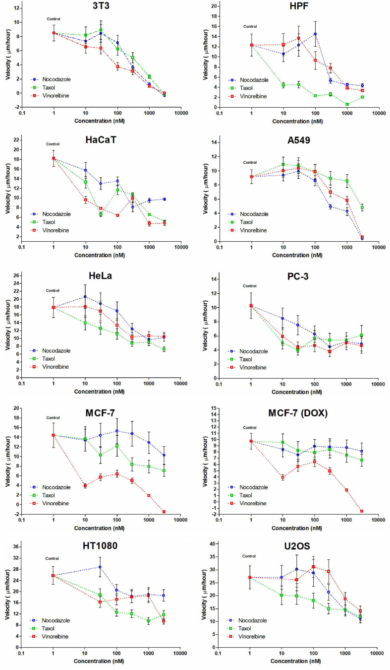
Wound healing assay performed with automated microscopy is widely used in drug testing, cancer cell analysis, and similar approaches. It is easy to perform, and the results are reproducible. However, it is usually used as a semi-quantitative approach because of inefficient image segmentation in transmitted light microscopy. Recently, several algorithms for wound healing quantification were suggested, but none of them was tested on a large dataset. In the current study, we develop a pipeline allowing to achieve correct segmentation of the wound edges in >95% of pictures and extended statistical data processing to eliminate errors of cell culture artifacts. Using this tool, we collected data on wound healing dynamics of 10 cell lines with 10 min time resolution. We determine that the overall kinetics of wound healing is non-linear; however, all cell lines demonstrate linear wound closure dynamics in a 6-h window between the fifth and 12th hours after scratching. We next analyzed microtubule-inhibiting drugs', nocodazole, vinorelbine, and Taxol, action on the kinetics of wound healing in the drug concentration-dependent way. Within this time window, the measurements of velocity of the cell edge allow the detection of statistically significant data when changes did not exceed 10-15%. All cell lines show decrease in the wound healing velocity at millimolar concentrations of microtubule inhibitors. However, dose-dependent response was cell line specific and drug specific. Cell motility was completely inhibited (edge velocity decreased 100%), while in others, it decreased only slightly (not more than 50%). Nanomolar doses (10-100 nM) of microtubule inhibitors in some cases even elevated cell motility. We speculate that anti-microtubule drugs might have specific effects on cell motility not related to the inhibition of the dynamic instability of microtubules.
利用自动显微镜进行的伤口愈合试验广泛应用于药物测试、癌细胞分析及类似研究中。该试验操作简便,结果具有可重复性。然而,由于透射光显微镜下图像分割效率低下,它通常被用作半定量方法。最近,有人提出了几种用于伤口愈合定量分析的算法,但均未在大型数据集上进行测试。在本研究中,我们开发了一种流程,能够在95%以上的图片中正确分割伤口边缘,并进行扩展的统计数据处理以消除细胞培养假象的误差。使用该工具,我们以10分钟的时间分辨率收集了10种细胞系的伤口愈合动力学数据。我们确定伤口愈合的总体动力学是非线性的;然而,所有细胞系在划痕后第5至12小时的6小时窗口内均表现出线性伤口闭合动力学。接下来,我们以药物浓度依赖的方式分析了微管抑制药物诺考达唑、长春瑞滨和紫杉醇对伤口愈合动力学的作用。在这个时间窗口内,当变化不超过10 - 15%时,细胞边缘速度的测量能够检测到具有统计学意义的数据。所有细胞系在毫摩尔浓度的微管抑制剂作用下,伤口愈合速度均降低。然而,剂量依赖性反应具有细胞系特异性和药物特异性。在某些细胞系中,细胞运动完全被抑制(边缘速度下降100%),而在其他细胞系中,仅略有下降(不超过50%)。在某些情况下,纳摩尔剂量(10 - 100 nM)的微管抑制剂甚至会提高细胞运动性。我们推测抗微管药物可能对细胞运动有特定影响,而与微管动态不稳定性的抑制无关。