Department of Medicine, Division of Pulmonary, Allergy & Critical Care Medicine, University of Alabama at Birmingham, Birmingham, AL, USA.
Program in Protease/Matrix Biology, University of Alabama at Birmingham, Birmingham, AL, USA.
Cell Rep Med. 2021 Apr 20;2(4):100242. doi: 10.1016/j.xcrm.2021.100242. Epub 2021 Mar 23.
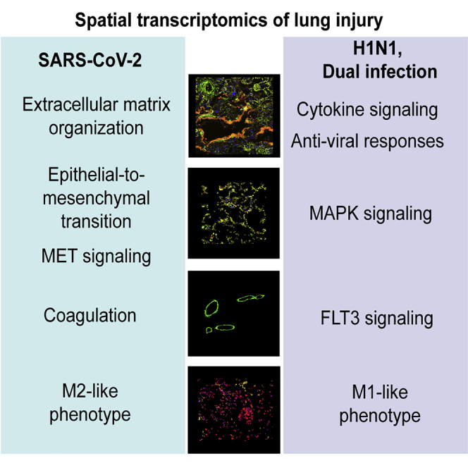
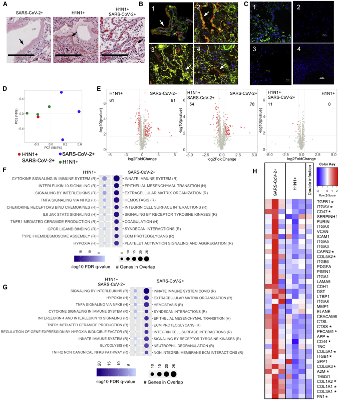
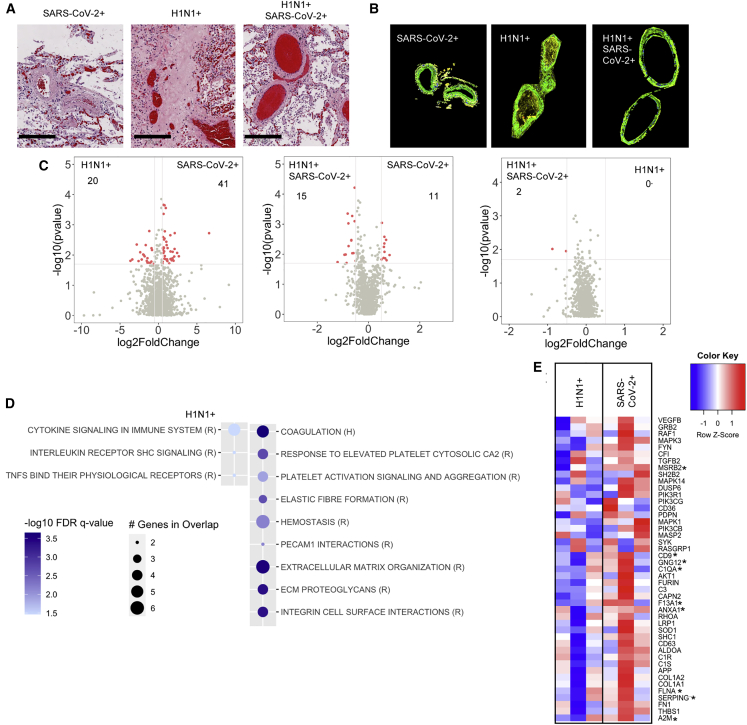
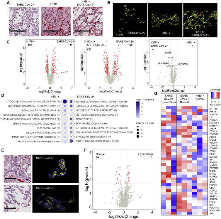
Severe SARS-CoV-2 infection often leads to the development of acute respiratory distress syndrome (ARDS), with profound pulmonary patho-histological changes post-mortem. It is not clear whether ARDS from SARS-CoV-2 is similar to that observed in influenza H1N1, another common viral cause of lung injury. Here, we analyze specific ARDS regions of interest utilizing a spatial transcriptomic platform on autopsy-derived lung tissue from patients with SARS-CoV-2 (n = 3), H1N1 (n = 3), and a dual infected individual (n = 1). Enhanced gene signatures in alveolar epithelium, vascular tissue, and lung macrophages identify not only increased regional coagulopathy but also increased extracellular remodeling, alternative macrophage activation, and squamous metaplasia of type II pneumocytes in SARS-CoV-2. Both the H1N1 and dual-infected transcriptome demonstrated an enhanced antiviral response compared to SARS-CoV-2. Our results uncover regional transcriptional changes related to tissue damage/remodeling, altered cellular phenotype, and vascular injury active in SARS-CoV-2 and present therapeutic targets for COVID-19-related ARDS.
严重的严重急性呼吸综合征冠状病毒 2 型(SARS-CoV-2)感染常导致急性呼吸窘迫综合征(ARDS),尸检后肺部病理变化明显。目前尚不清楚 SARS-CoV-2 引起的 ARDS 是否与流感 H1N1 所致的 ARDS 相似,流感 H1N1 也是肺部损伤的常见病毒性病因之一。在此,我们利用 SARS-CoV-2(n=3)、H1N1(n=3)和 1 例双重感染患者的尸检肺组织,通过空间转录组学平台分析特定的 ARDS 感兴趣区域。在 SARS-CoV-2 中,肺泡上皮、血管组织和肺巨噬细胞中增强的基因特征不仅表明局部凝血功能障碍增加,而且表明细胞外重塑、替代型巨噬细胞激活和 II 型肺泡上皮细胞的鳞状化生增加。与 SARS-CoV-2 相比,H1N1 和双重感染的转录组均显示出增强的抗病毒反应。我们的研究结果揭示了与组织损伤/重塑、细胞表型改变和血管损伤相关的区域性转录变化,这些变化在 SARS-CoV-2 中活跃,并为 COVID-19 相关 ARDS 提供了治疗靶点。