Department of Radiology, Vrije Universiteit Brussel (VUB), Universitair Ziekenhuis Brussel (UZ Brussel), Laarbeeklaan 101, 1090, Brussels, Belgium.
Department of Oncology, University of Oxford, Headington, Oxford, OX3 7LE, UK.
Eur Radiol. 2021 Oct;31(10):7540-7549. doi: 10.1007/s00330-021-07763-7. Epub 2021 Mar 30.
Routine dosimetry calculations do not account for the presence of iodine in organs and tissues during CT acquisition. This study aims to investigate the impact of contrast agent (CA) on radiation dose.
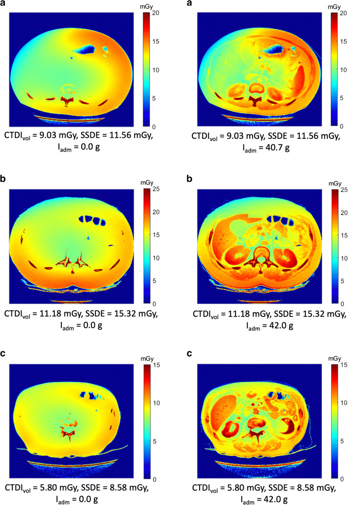
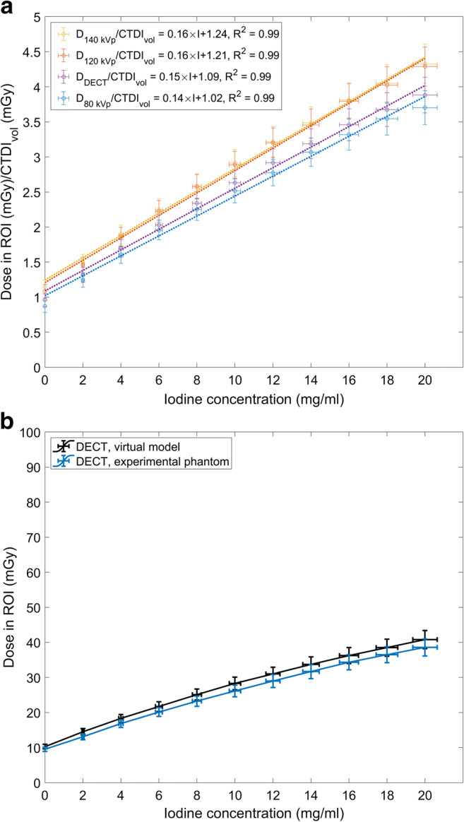
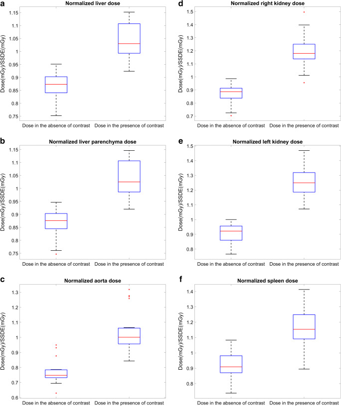
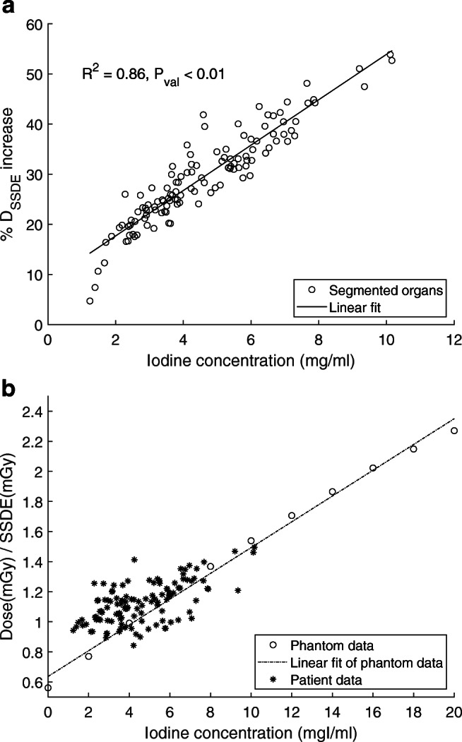
First, relation between absorbed radiation dose and iodine concentrations was investigated using a cylindrical water phantom with iodine-saline dilution insertions. Subsequently, a retrospective study on abdominal dual-energy CT (DECT) patient data was performed to assess the increase of the local absorbed radiation dose compared to a non-contrast scan. Absorbed doses were estimated with Monte Carlo simulations using the individual CT voxel data of phantom and patients. Further, organ segmentations were performed to obtain the dose in liver, liver parenchyma, left kidney, right kidney, aorta, and spleen.
In the phantom study, a linear relation was observed between the radiation dose normalized by computed tomography dose index (CTDI) and CA concentrations I (mg/ml) for three tube voltages; [Formula: see text] = 0.14 × I + 1.02, [Formula: see text] = 0.16 × I + 1.21, [Formula: see text] = 0.16 × I + 1.24, and for DECT acquisition; [Formula: see text] = 0.15 × I + 1.09. Similarly, a linear relation was observed between the dose increase and the organ iodine contents (R = 0.86 and p < 0.01) in the patient study. The relative doses increased in the liver (21 ± 5%), liver parenchyma (20 ± 5%), right kidney (37 ± 7%), left kidney (39 ± 7%), aorta (34 ± 6%) and spleen (26 ± 4%). In addition, the local dose distributions changed based on patient's anatomy and physiology.
Compared to a non-contrast scan, the organ doses increase by 30% in contrast-enhanced abdominal CT. This study suggests considering CA in dosimetry calculations, epidemiological studies, and organ dose estimations while developing new CT protocols.
• The presence of contrast media increases radiation absorption in CT, and this increase is related to the iodine content in the organs. • The increased radiation absorption due to contrast media can lead to an average 30% increase in absorbed organ dose. • Iodine should be considered in CT radiation safety studies.
在 CT 采集过程中,常规剂量计算未考虑器官和组织中碘的存在。本研究旨在探讨对比剂(CA)对辐射剂量的影响。
首先,使用带有碘盐水稀释插入物的圆柱形水模体研究吸收辐射剂量与碘浓度之间的关系。随后,对腹部双能 CT(DECT)患者数据进行回顾性研究,以评估与非对比扫描相比,局部吸收辐射剂量的增加。使用蒙特卡罗模拟根据模体和患者的个体 CT 体素数据估算吸收剂量。此外,还进行了器官分割,以获得肝脏、肝实质、左肾、右肾、主动脉和脾脏的剂量。
在体模研究中,对于三种管电压,观察到经 CT 剂量指数(CTDI)归一化的辐射剂量与 CA 浓度 I(mg/ml)之间呈线性关系;[公式:见正文] = 0.14 × I + 1.02,[公式:见正文] = 0.16 × I + 1.21,[公式:见正文] = 0.16 × I + 1.24,对于 DECT 采集;[公式:见正文] = 0.15 × I + 1.09。同样,在患者研究中,观察到剂量增加与器官碘含量之间存在线性关系(R = 0.86,p < 0.01)。在肝脏(21 ± 5%)、肝实质(20 ± 5%)、右肾(37 ± 7%)、左肾(39 ± 7%)、主动脉(34 ± 6%)和脾脏(26 ± 4%)中,相对剂量增加。此外,基于患者的解剖结构和生理学,局部剂量分布发生变化。
与非对比扫描相比,腹部 CT 增强扫描时器官剂量增加 30%。本研究表明,在开发新的 CT 方案时,应考虑对比剂在剂量计算、流行病学研究和器官剂量估算中的作用。
对比剂的存在会增加 CT 中的辐射吸收,这种增加与器官中的碘含量有关。
由于对比剂引起的辐射吸收增加可导致吸收器官剂量平均增加 30%。
在 CT 辐射安全研究中应考虑碘。